Learn More
Smad2 (pS465/pS467)/Smad3 (pS423/pS425) Mouse, PE, Clone: O72-670, BD
Shop All BD Biosciences ProductsDescription
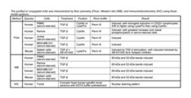
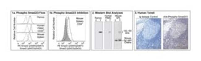
The O72-670 monoclonal antibody specifically binds to the Smad2 protein phosphorylated at the Ser465/467 sites and the Smad3 protein phosphorylated at the Ser423/425 sites. Smad2 and Smad3 are members of the Smad superfamily with observed molecular weights of 60kDa and 52kDa, respectively. The Smad family consists of three subfamilies: receptor regulated Smads or R-Smads, including Smads1, 2, 3, 5, and 8; common partner Smad, or Co-Smad, including Smad4; and inhibitory Smads, or I-Smad, including Smads 6 and 7. Activation of TGF-beta superfamily serine/threonine kinase receptors, such as TGF-beta, activin and BMP receptors, by their bound ligands leads to the phosphorylation of R-Smads at several sites. It has been shown that the ligand-bound TGF-beta type I receptor directly phosphorylates Ser465 and Ser467 of Smad2 and Ser423 and Ser425 of Smad3. Phosphorylated R-Smads form complexes with Co-Smad and translocate into the nucleus to regulate transcription affecting a wide range of critical cellular processes including cell-fate determination, proliferation, morphogenesis, differentiation and apoptosis. The inhibitory Smads inhibit this pathway through two potential mechanisms: either by preventing R-Smads from binding to their corresponding receptors and/or by competing with Smad4, the Co-Smad, from binding to R-Smads. High level expression of phosphorylated Smad2 has been associated with poor prognosis in late stage gastric carcinoma. Roles for Smad2 have been described in thymopoiesis and the TGF-β-mediated induction of regulatory T cells and Th17 cells. The specificity of the O72-670 mAb was confirmed by Western blot and immunohistochemistry using unconjugated antibody.
Host Species: Mouse
Clone: O72-670
Isotype: IgG1, κ
Species Reactivity: Human
Immunogen: Phosphorylated Human Smad2 Peptide
Intracellular Staining
Specifications
Specifications
| Antigen | Smad2 (pS465/pS467)/Smad3 (pS423/pS425) |
| Applications | Flow Cytometry |
| Classification | Monoclonal |
| Clone | O72-670 |
| Conjugate | PE |
| Description | SMAD2, SMAD3; MADH2, MADH3; MAD homolog 2, MAD homolog 3 |
| Formulation | Aqueous buffered solution containing BSA and ≤0.09% sodium azide. |
| Host Species | Mouse |
| Immunogen | Phosphorylated Human Smad2 Peptide |
| Purification Method | Affinity Purified |
| Show More |
By clicking Submit, you acknowledge that you may be contacted by Fisher Scientific in regards to the feedback you have provided in this form. We will not share your information for any other purposes. All contact information provided shall also be maintained in accordance with our Privacy Policy.