Learn More
Description
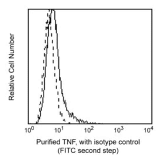
Clone MAb11 reacts with the human form of tumor necrosis factor (TNF, formerly known as TNF-α) detectable in the cytoplasm of a major subset of activated peripheral blood T lymphocytes. Using standard procedures for detection of intracellular proteins, clone MAb11 also cross-reacts with activated peripheral blood CD3+ lymphocytes of baboon, and both rhesus and cynomolgus macaque monkeys following five-hour treatment with phorbol myristic acetate (PMA) and Ca++ Ionophore (A23187) in the presence of monensin. The staining is restricted to CD3+ T cells and is similar to that observed with peripheral blood T lymphocytes from normal human donors. This antibody is routinely tested by flow cytometric analysis. Other applications were tested at BD Biosciences Pharmingen during antibody development only or reported in the literature.
Intracellular Staining
Specifications
Specifications
| Antigen | TNF |
| Applications | Blocking Assay, Flow Cytometry |
| Classification | Monoclonal |
| Clone | MAB11 |
| Concentration | 0.5mg/mL |
| Conjugate | Unconjugated |
| Formulation | Aqueous buffered solution containing ≤0.09% sodium azide. |
| Host Species | Mouse |
| Immunogen | Recombinant Human TNF |
| Purification Method | Affinity Purified |
| Show More |
By clicking Submit, you acknowledge that you may be contacted by Fisher Scientific in regards to the feedback you have provided in this form. We will not share your information for any other purposes. All contact information provided shall also be maintained in accordance with our Privacy Policy.