Learn More

Invitrogen™ beta Amyloid Polyclonal Antibody (CT695)


Description
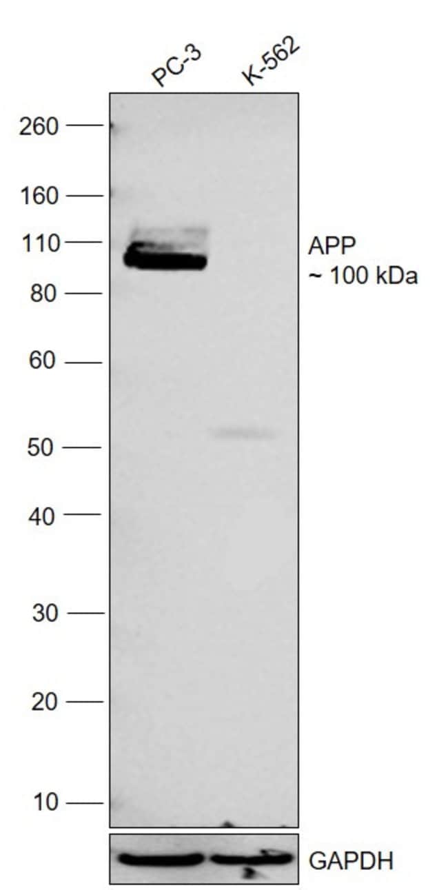
This antibody can be used to specifically detect the beta-amyloid precursor protein. The antibody reacts with full-length (APP695, 751, 770) and N-terminal truncated forms of beta-APP. The antibody can also be used to detect the C-terminal membrane-anchored fragment of beta-APP that remains after alpha- or beta-secretase cleavage. This antibody does not detect the beta-APP product N-terminal to the gamma-secretase cleavage site. Reactivity with this antibody has been confirmed for human, mouse, pig, and rat. Based on sequence homology, reactivity with other species, including monkey, guinea pig, and chicken beta-APP, is highly likely. The CT695 antibody has demonstrated superiority in detecting axonal damage. This antibody has been tested in western blotting with FRTL-5 (rat thyroid) cells and rat brain lysates. For immunohistochemistry FFPE tissues will require epitope retrieval pretreatment.
Specifications
Specifications
| Antigen | beta Amyloid |
| Applications | ELISA, Immunohistochemistry, Western Blot |
| Classification | Polyclonal |
| Clone | CT695 |
| Concentration | 0.25 mg/mL |
| Conjugate | Unconjugated |
| Formulation | PBS with 0.1% sodium azide; pH 7.4 |
| Gene | APP |
| Gene Accession No. | P05067, P08592, P12023, P79307 |
| Gene Alias | A4; AAA; ABETA; Abeta40; Abeta42; ABPP; AD1; Adap; AG; AICD-50; AICD-57; AICD-59; AID(50); AID(57); AID(59); Alpha-CTF; Alpha-secretase C-terminal fragment; Alzheimer disease; Alzheimer disease amyloid A4 protein homolog; alzheimer disease amyloid protein; Amyloid; amyloid A4; Amyloid b; Amyloid beta; amyloid beta (A4) precursor protein; amyloid beta (A4) precursor protein (peptidase nexin-II, Alzheimer disease); amyloid beta A4 protein; amyloid beta precursor protein; Amyloid intracellular domain 50; Amyloid intracellular domain 57; Amyloid intracellular domain 59; Amyloid precursor protein; amyloid precursor protein variant 1; amyloid precursor protein variant 2; Amyloid β; amyloid-beta A4 protein; amyloid-beta A4 protein; amyloid beta A4 protein; Amyloid-beta precursor protein; Amyloid-beta protein 40; Amyloid-beta protein 42; Amyloidogenic glycoprotein; APP; APP-C57; APP-C59; APP-C99; APPI; appican; beta amyloid protein; beta amyloid protein precursor; beta-amyloid peptide; beta-amyloid peptide(1-40); beta-amyloid peptide(1-42); Beta-amyloid precursor protein; betaApp; Beta-APP40; Beta-APP42; Beta-CTF; Beta-secretase C-terminal fragment; C31; C80; C83; C99; Cerebral vascular amyloid peptide; CTF gamma; CTF-alpha; CTFgamma; CVAP; E030013M08Rik; Gamma-CTF(50); Gamma-CTF(57); Gamma-CTF(59); Gamma-secretase C-terminal fragment 50; Gamma-secretase C-terminal fragment 57; Gamma-secretase C-terminal fragment 59; N-APP; OTTHUMP00000096096; P3(40); P3(42); Pan-Abeta; peptidase nexin-II; PN2; PN-II; PreA4; PreA4 751; protease nexin II; protease nexin-II; S-APP-alpha; S-APP-beta; Soluble APP-alpha; Soluble APP-beta; testicular tissue protein Li 2 |
| Show More |
Frequently Asked Questions (FAQs)
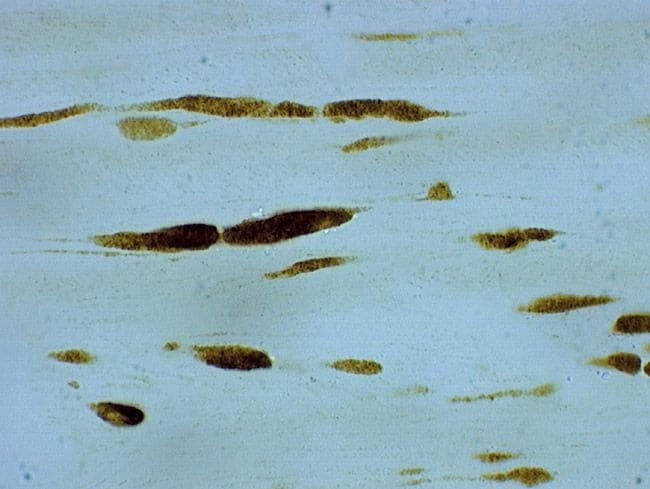
Here is a procedure for the immunohistochemical staining of Beta-amyloid with paraffin embedded sections of transgenic mouse brain. The following protocol was developed for use with paraffin embedded sections stained with a variety of our antibodies against Beta-amyloid. Applicable rabbit and mouse anti-Beta-amyloid antibodies are: 51-2700, 13-0200, 71-5800, 13-0100Z, 37-4200, 43-7900, 44-136, 700254, 36-6900, and AHB0121.
Beta-amyloid Staining:
- Transgenic mice (expressing the transgenes PS1-A246E + APP swe, APP swe alone, or PS1-A246E alone) were perfused with 1 x Dulbecco's phosphate buffered saline (D-PBS) followed by 4% paraformldehyde buffered with D-PBS. Brain tissues were then embedded in paraffin prior to microtome sectioning.
- After mounting on slides, the paraffin-embedded tissue sections were then deparaffinized with heat. The sections were then incubated for 3 minutes in 70% formic acid. Next, they were deparaffinized further with xylene followed with 100% ethanol. The sections were then re-hydrated in a graded ethanol series (100% ethanol, 95% ethanol, 70% ethanol, and then water).
- Endogenous peroxidase activity was quenched with 3% hydrogen peroxide in methanol. The sections were then heated in the microwave for 5-7 minutes in water, cooled at room temperature for 5 minutes and then rinsed in water. The sections were then washed in TBS (0.05 M Tris-HCl, pH 7.6, with 0.25 M NaCl) prior to blocking. Non-specific binding was blocked with 3% normal goat serum and 0.1% Triton X-100 in TBS for 1 hour at room temperature.
- The sections were then stained with anti-Beta-amyloid antibodies. The staining solution typically consisted of the antibody at a concentration of 5 µg/mL in TBS containing 2% normal goat serum. After incubation at room temperature for at least 1 hr., the sections were washed in TBS 3 times for 5 minutes each. An anti-mouse or anti-rabbit secondary antibody labeled with HRP was then used with DAB or AEC as substrates to stain the Beta-amyloid.
[Adapted from Borchelt, D.R., et al. (1997) Accelerated amyloid deposition in the brains of transgenic mice coexpressing mutant presenilin 1 and amyloid precursor proteins. Neuron 19:939-945.]
Tau and Synuclein Staining:
Here is a general procedure for the immunohistochemical staining of tau, its phosphorylated forms, and synuclein in frozen sections of rat brain. This protocol is adapted from one kindly contributed by Dr. Emil Adamec, M.D., Ph.D., McLean Hospital, Belmont, MA. The procedure was developed for use with cryostat sections of rat brain.
- Sections were fixed with 4% paraformaldehyde for 25 minutes. Sections were then extracted with 0.01% (v/v) Triton X-100. Endogenous peroxidase was blocked with 3% hydrogen peroxide. Sections were then treated with 80% formic acid for 5 to 10 minutes at room temperature to enhance staining. Note that formic acid treatment is also very useful prior to staining with anti-synuclein antibodies.
- The various antibodies were used at dilutions of 1:100 to 1:250. The sections were incubated with the diluted primary antibody overnight at 4°C. A species-specific secondary antibody labeled with HRP was then used with either DAB or AEC to stain the antigens.
- Applicable rabbit and mouse anti-tau and phospho-tau antibodies are: AHB0042, AHB0061, 44-738G, 39-1800, 13-6400, 13-1400, and 18-7461. Anti-synuclein antibodies useful for IHC are: 18-0215, 18-7461, 32-8100, 32-8200, 32-8500, 35-8300, 35-8400, 39-1800, and AHB0261.
We recommend that hydrophobic residues comprise 50% or less of all the residues in your sequence. Make sure there is at least one charged residue for every five amino acids: this is generally known to enhance the solubility of the peptide. Peptides (compared to polypeptides, which fold and bury the hydrophobic amino acids) are too small to fold, so just a few hydrophobic amino acids may leave them insoluble.
Other guidelines include:
Peptides containing multiple Cys, Met, and Trp can be hard to synthesize.
Some sequences are problematic in solid-phase peptide synthesis or cleavage and are best avoided; these include Asp-Pro sequences or stretches of amino acids that require bulky protecting groups on their side chains during synthesis.
Glycine is often good for antigenicity as it has only a hydrogen side chain; this allows for complete rotation.
Please note that we offer a Custom Antibody Production service (https://www.thermofisher.com/us/en/home/life-science/antibodies/custom-antibodies/custom-antibody-production.html) that includes the use of our proprietary Antigen Profiler and Antigen Preparation tool (https://www.thermofisher.com/us/en/home/life-science/antibodies/custom-antibodies/custom-antibody-production/antigen-profiler-antigen-preparation.html).
Most peptide antigens range in length from 12 to 16 residues and are relatively easy to synthesize. Peptides of 9 residues or shorter have been effective antigens for antibody production, but peptides longer than 16 amino acids may contain several epitopes and form secondary structures. Peptides in excess of 18 residues begin to present more synthetic challenges. Before you synthesize your peptide, we recommend doing a BLASTP search using your peptide sequence. This is to make sure that the peptide is not homologous or identical to a sequence in a completely unrelated protein in the host animal.
Please note that we offer a Custom Antibody Production service (https://www.thermofisher.com/us/en/home/life-science/antibodies/custom-antibodies/custom-antibody-production.html) that includes the use of our proprietary Antigen Profiler and Antigen Preparation tool (https://www.thermofisher.com/us/en/home/life-science/antibodies/custom-antibodies/custom-antibody-production/antigen-profiler-antigen-preparation.html).
Improper storage of antibodies can lead to:
- Degradation: Loss of activity and specificity
- Aggregation: Formation of precipitates or clumps
- Reduced performance: Poor results in assays or experiments
Antibody pair kits contain capture antibody, detection antibody, recombinant standard and HRP conjugate. Each contains enough reagents to process forty 96-well plates. A list of Antibody Pair Kits (https://www.thermofisher.com/us/en/home/life-science/protein-biology/protein-assays-analysis/elisa/antibody-pair-kits.html) is available by target.
Safety and Handling
By clicking Submit, you acknowledge that you may be contacted by Fisher Scientific in regards to the feedback you have provided in this form. We will not share your information for any other purposes. All contact information provided shall also be maintained in accordance with our Privacy Policy.