Promotional price valid on web orders only. Your contract pricing may differ. Interested in signing up for a dedicated account number?
Learn More

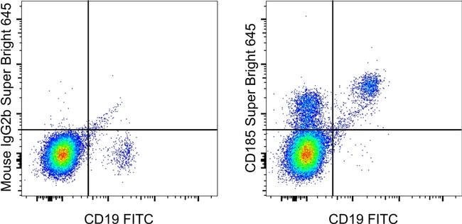
Invitrogen™ CD185 (CXCR5) Monoclonal Antibody (MU5UBEE), Super Bright™ 645, eBioscience™, Invitrogen™
GREENER_CHOICE

Catalog No. 64918542
Encompass_Preferred
Change view
Click to view available options
Quantity:
100 Tests
1 product options available for selection
Product selection table with 1 available options. Use arrow keys to navigate and Enter or Space to select.
Catalog No. Quantity
64-918-542 100 Tests
Use arrow keys to navigate between rows. Press Enter or Space to select a product option. 1 options available.
1 options
Catalog No. 64-918-542 Supplier Invitrogen™ Supplier No. 64918542
Add to Cart
Add to Cart

Mouse Monoclonal Antibody

Description: The RMST2-2 monoclonal antibody reacts with ST2, also known as IL-33 receptor (IL-33R) or interleukin 1 receptor-like 1. ST2 is a member of the IL-1 receptor family. In the membrane-bound form, it consists of three extra-cellular immunoglobulin domains and an intracellular toll-interleukin-1 receptor domain. ST2 forms a dimer with IL-1R accessory protein in a ligand-dependent manner. The IL-33/ST2 interaction induces the production of TH2 cytokines. ST2 also has a soluble isoform lacking transmembrane and intracellular toll-interleukin-1 receptor domains. It is believed that the soluble form functions as a decoy receptor that can block membrane bound IL-33/ST2 interaction. ST2 is expressed by TH2 lymphocytes, mast cells, eosinophils, basophils, innate lymphocytes, smooth muscle cells, and endothelial cells and is involved in host defense, allergy, and inflammation. The IL-33/ST2 interaction has also been shown to be atheroprotective. Applications Reported: This RMST2-2 antibody has been reported for use in flow cytometric analysis. Applications Tested: This RMST2-2 antibody has been tested by flow cytometric analysis. This may be used at less than or equal to 1.0 μg per test. A test is defined as the amount (μg) of antibody that will stain a cell sample in a final volume of 100 μL. Cell number should be determined empirically but can range from 10^5 to 10^8 cells/test.

CD185, also known as CXCR5 or Burkitt lymphoma receptor 1 (BLR1), is a seven-transmembrane G protein-coupled receptor in the CXC chemokine receptor family. It plays a crucial role in lymphocyte trafficking, particularly in T cell migration into B cell follicles of germinal centers in response to CXCL13, making it a key marker of follicular helper T cells. In peripheral blood, CXCR5 is expressed on B cells, CD4+ T cells (excluding Th1 and Th2 cells), and a subset of memory (CD45RO+) T cells. These CD185+ T cells are in a resting state and migrate to lymph nodes due to CCR7 and CD62L expression. In tonsils, CD185 is found on nearly all CD4+ cells, along with activation markers like CD69 and ICOS, and can induce antibody production when co-cultured with B cells. CXCR5 is expressed in lymphatic tissues such as lymph node follicles and the spleen, and is also found in human blood and salivary glands, as well as various animal tissues. It is essential for B cell migration and is part of the larger family of G protein-coupled receptors (GPCRs), which have seven membrane-spanning helices and are involved in numerous signaling pathways. GPCRs, including CXCR5, are regulated by various ligands that affect their conformation and signaling through G-proteins and downstream effectors. The regulation of GPCRs, including their deactivation, has significant implications for cellular functions and is a major focus of pharmacological research, leading to the development of drugs like Zyprexa and Claritin for mental health and allergy treatments.
TRUSTED_SUSTAINABILITY

Specifications

Antigen CD185 (CXCR5)
Applications Flow Cytometry
Classification Monoclonal
Clone MU5UBEE
Concentration 5 μL/Test
Conjugate Super Bright 645
Formulation PBS with BSA and 0.09% sodium azide; pH 7.2
Gene CXCR5
Gene Accession No. P32302, Q04683
Gene Alias Blr1; Blr-1; Burkitt lymphoma receptor 1; burkitt lymphoma receptor 1 homolog; Burkitt lymphoma receptor 1, GTP binding protein (chemokine (C-X-C motif) receptor 5); Burkitt lymphoma receptor 1, GTP-binding protein; C Cmotif chemokine; C X C motif chemokine; CC motif chemokine; CCmotif chemokine; CD185; CD185 antigen; chemochine (C-X-C motif) receptor 5; chemokine (C-X-C motif) receptor 5; CXC; C-X-C chemokine receptor type 5; CXC motif chemokine; C-X-C motif chemokine receptor 5; CXCR5; CXC-R5; CXCR-5; EGK_06966; Fusin; Gpcr6; GPR9; HUMSTR; LESTR; MDR15; MDR-15; Monocyte-derived receptor 15; neurolymphatic receptor; NLR
Gene Symbols CXCR5
Host Species Mouse
Immunogen N-terminal extracellular domain.
Purification Method Affinity chromatography
Quantity 100 Tests
Regulatory Status RUO
Primary or Secondary Primary
Gene ID (Entrez) 12145, 643, 701792
Target Species Human, Mouse, Rhesus Macaque
Content And Storage 4°C, store in dark, DO NOT FREEZE!
Product Type Antibody
Form Liquid
Isotype IgG2b κ
Show More Show Less
Can I use the OneComp and UltraComp eBeads microspheres with Super Bright-conjugated antibodies?

UltraComp eBeads microspheres (Cat. No. 01-2222) are recommended for use with Super Bright dyes.
Note: Super Bright Staining Buffer (Cat. No. SB-4400) is not compatible with UltraComp eBeads microspheres (Cat. No. 01-2222-41, 00-2222-42). If using UltraComp eBeads microspheres as a compensation tool, solely use Flow Cytometry Stain Buffer (Cat. No. 00-4222-26, 00-4222-57) for any antibody dilutions.

In some experiments, we have observed that compensation values for Super Bright 780- and Brilliant Violet 785- or Brilliant Violet 786-conjugated antibodies are higher in the violet 450/50 channel when using UltraComp eBeads microspheres as compared to single-color stained cells. In such circumstances, we would recommend setting compensation with cells. We have also observed this in some experiments using AbC Total Antibody Compensation beads, both with Super Bright 780 and Brilliant Violet 786. We have not tested Brilliant Violet 785 with the AbC beads.

Can I prepare an antibody cocktail containing Super Bright Staining Buffer and Super Bright-conjugated antibodies ahead of time?

We recommend that the antibody cocktails containing Super Bright-conjugated antibodies and Super Bright Staining Buffer are prepared fresh prior to staining. Discard any unused portions. We do not recommend overnight storage of prepared cocktails.

Can I fix my cells after staining with Super Bright-conjugated antibodies? How long can fixed cells be stored prior to analysis?

Samples that have been stained with antibodies conjugated to Super Bright dyes may be stored for up to three days, at 2-8°C, in the dark, using either IC Fixation Buffer (Cat. No. 00-8222) or 1-step Fix/Lyse Buffer (Cat. No. 00-5333) with no significant effect on brightness or compensation.

Are the Super Bright dyes sensitive to methanol fixation?

Super Bright dyes are stable in methanol-based fixation buffers.

Can Super Bright-conjugated antibodies be used in combination with intracellular (IC Fixation/Permeabilization) or intranuclear (Foxp3 fixation/permeabilization) staining?

Yes, Super Bright-conjugated antibodies are stable in formaldehyde-based fixation buffers and permeabilization buffers, such as the IC Fixation and Permeabilization Buffer Set (Cat. No. 88-8824) and the Foxp3/Transcription Factor Staining Buffer Set (Cat. No. 00-5523).

Which laser do I use to detect Super Bright-conjugated antibodies?

The violet laser (405 nm) should be used to excite Super Bright-conjugated antibodies.

Can the Super Bright Staining Buffer be used with other polymer dyes?

Yes, the Super Bright Staining Buffer (Cat. No. SB-4400) is compatible with other polymer dyes (i.e., Brilliant Violet dyes) and is useful for minimizing any non-specific polymer interactions when two or more of these dyes are used in combination.

Can Super Bright-conjugated antibodies be used in combination with other polymer dyes? What buffer should I use when using more than one Super Bright or polymer dye?

When using two or more Super Bright dyes, or when using Super Bright dyes in combination with other polymer dyes (i.e., Brilliant Violet dyes), we recommend using the Super Bright Staining Buffer (Cat. No. SB-4400) to minimize any non-specific polymer interactions between these fluorochromes. For specific instructions for use, please refer to the product technical data sheet.

Are there recommended tubes for staining with Super Bright-conjugated antibodies?

We recommend using polystyrene tubes (for example, FACS tubes) for staining with your Super Bright-conjugated antibodies. If using polypropylene (for example, Eppendorf tubes), protecting from light is critical.

Is there a specific buffer I should use when staining with Super Bright-conjugated antibodies?

No special buffer is required when using a single Super Bright-conjugated antibody in a panel.
When using more than one Super Bright dye, or when using Super Bright dyes in combination with other polymer dyes (i.e., Brilliant Violet dyes), we recommend using the Super Bright Staining Buffer (Cat. No. SB-4400) to minimize any non-specific polymer interactions between these fluorochromes. For specific instructions for use, please refer to the product technical data sheet.

How does the protocol for staining with Super Bright-conjugated antibodies differ from staining with conventional organic fluorochrome and eVolve conjugated antibodies?

Antibodies conjugated to Super Bright dyes can be used similarly to traditional fluorochromes. If multiple Super Bright dyes are used in combination with each other or in combination with other polymer dyes (i.e., Brilliant Violet dyes), then the use of Super Bright Staining Buffer (Cat. No. SB-4400) is recommended to minimize any non-specific polymer interactions between these fluorochromes. For specific instructions for use, please refer to the product technical data sheet.

Can the Super Bright dyes be used for non-flow applications?

Super Bright dyes have not been tested for applications other than flow cytometry.

What is the difference between the Super Bright and eVolve dyes?

Super Bright dyes are fluorochromes based on polymer technology and are excited by the violet laser. In contrast, eVolve dyes are based on Qdot nanocrystal technology and are maximally excited by the UV laser, although they may also be excited by violet, blue, yellow-green, and, depending on the eVolve, the red laser lines. The general shape of the emission spectra for Super Bright dyes will be more similar to traditional fluorochromes, while eVolve dyes have extremely narrow emission spectra. Please contact Tech Support (techsupport@thermofisher.com) for more information.

What are the Super Bright dyes?

eBioscience Super Bright dyes are a series of patent-pending fluorochromes that are based upon a fluorescent polymer and its tandems. Super Bright dyes have been developed for use in flow cytometry and can be excited by the violet laser (405 nm). The peak emission of each Super Bright dye is indicated by the number in the product description, e.g., "Super Bright 600" has a peak emission of 600 nm.

Are the Super Bright Dyes photo-labile?

As with other fluorochromes, we recommend minimal exposure to light to maintain optimal signal.


Product Title
Select an issue

By clicking Submit, you acknowledge that you may be contacted by Fisher Scientific in regards to the feedback you have provided in this form. We will not share your information for any other purposes. All contact information provided shall also be maintained in accordance with our Privacy Policy.