Learn More
Invitrogen™ CD1a Monoclonal Antibody (HI149), PerCP-eFluor™ 710, eBioscience™


Description
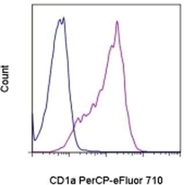
Description: The HI149 monoclonal antibody reacts with human CD1a, a 49 kDa protein expressed by cortical thymocytes and dendritic cells including Langerhans cells. The CD1 family of proteins share some structural and functional characteristics with the MHC class I molecules; however, members of the CD1 family are not polymorphic. Similar to MHC class I, CD1a associates with the beta2-microglobulin and is thought to play a role in antigen presentation. Applications Reported: This HI149 antibody has been reported for use in flow cytometric analysis. Applications Tested: This HI149 antibody has been pre-titrated and tested by flow cytometric analysis of the MOLT-4 cell line. This can be used at 5 μL (0.25 μg) per test. A test is defined as the amount (μg) of antibody that will stain a cell sample in a final volume of 100 μL. Cell number should be determined empirically but can range from 10^5 to 10^8 cells/test. PerCP-eFluor® 710 emits at 710 nm and is excited with the blue laser (488 nm); it can be used in place of PerCP-Cyanine5.5. We recommend using a 710/50 bandpass filter, however, the 695/40 bandpass filter is an acceptable alternative. Please make sure that your instrument is capable of detecting this fluorochrome. Fixation: Samples can be stored in IC Fixation Buffer (Product # 00-822-49) (100 μL cell sample + 100 μL IC Fixation Buffer) or 1-step Fix/Lyse Solution (Product # 00-5333-54) for up to 3 days in the dark at 4°C with minimal impact...
Specifications
Specifications
| Antigen | CD1a |
| Applications | Flow Cytometry |
| Classification | Monoclonal |
| Clone | HI149 |
| Concentration | 5 μL/Test |
| Conjugate | PerCP-eFluor 710 |
| Formulation | PBS with BSA and 0.09% sodium azide; pH 7.2 |
| Gene | CD1A |
| Gene Accession No. | P06126 |
| Gene Alias | CD1; CD1 antigen; CD1.1; CD1A; CD1A antigen, a polypeptide; CD1a molecule; cd1a1; CD1A1 antigen; cluster of differentiation 1 A; cortical thymocyte antigen CD1A; differentiation antigen CD1-alpha-3; epidermal dendritic cell marker CD1a; FCB6; HTA1; hTa1 thymocyte antigen; R4; T6; T-cell surface antigen T6/Leu-6; T-cell surface glycoprotein CD1a |
| Show More |
Frequently Asked Questions (FAQs)
The eFluor Organic fluorochromes and eVolve QDots can be used with flow staining buffers containing PBS and protein.
The eFluor Organic Dyes (eFluor 450, APC-eFluor 780, PerCP-eFluor 710, eFluor 710) are conventional fluorochromes. In contrast, the eVolve line of products are Quantum dots.
As with other fluorochromes, we recommend minimal exposure to light to maintain optimal signal.
Safety and Handling
By clicking Submit, you acknowledge that you may be contacted by Fisher Scientific in regards to the feedback you have provided in this form. We will not share your information for any other purposes. All contact information provided shall also be maintained in accordance with our Privacy Policy.