Learn More
Invitrogen™ CD1c Monoclonal Antibody (L161), PerCP-eFluor™ 710, eBioscience™, Invitrogen™

Description
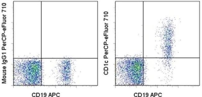
Description: This L161 monoclonal antibody detects CD1c (also known as BDCA-1), a glycoprotein that is noncovalently linked to beta-2 microglobulin on thymocytes and antigen presenting cells such as dendritic and Langerhans cells. This molecule is also expressed on some circulating and marginal zone B cells, as well as in lymph nodes and germinal centers. CD1c is involved in the presentation of lipid antigens such as microbial fatty acids to effector T cells during the adaptive immune response. Finally, alternative splicing gives rise to three different isoforms of CD1c (soluble, membrane, and cytoplasmic/soluble isoforms). Applications Reported: This L161 antibody has been reported for use in flow cytometric analysis. Applications Tested: This L161 antibody has been pre-titrated and tested by flow cytometric analysis of normal human peripheral blood cells. This can be used at 5 μL (0.06 μg) per test. A test is defined as the amount (μg) of antibody that will stain a cell sample in a final volume of 100 μL. Cell number should be determined empirically but can range from 10^5 to 10^8 cells/test. PerCP-eFluor™ 710 emits at 710 nm and is excited with the blue laser (488 nm); it can be used in place of PerCP-Cyanine5.5. We recommend using a 710/50 bandpass filter, however, the 695/40 bandpass filter is an acceptable alternative. Please make sure that your instrument is capable of detecting this fluorochrome.
Specifications
Specifications
| Antigen | CD1c |
| Applications | Flow Cytometry |
| Classification | Monoclonal |
| Clone | L161 |
| Concentration | 5 μL/Test |
| Conjugate | PerCP-eFluor 710 |
| Formulation | PBS with BSA and 0.09% sodium azide; pH 7.2 |
| Gene | CD1C |
| Gene Accession No. | P29017 |
| Gene Alias | BDCA1; canCD1c; CD1; CD1A; CD1C; CD1C antigen, c polypeptide; CD1c molecule; cortical thymocyte antigen CD1C; differentiation antigen CD1-alpha-3; R7; RP11-101J8.3; T-cell surface glycoprotein CD1c |
| Show More |
Frequently Asked Questions (FAQs)
Our options will depend on the samples you are analyzing.
If cell viability is not critical, you can store your stained samples at 4 degrees C or on ice overnight in the dark and analyze the following day.
For samples stained with eFluor organic fluorochromes, we recommend that cells be suspended in 100 uL of Flow Cytometry Staining Buffer (Cat. No. 00-4222) and 100 uL of eBioscience IC Fixation Buffer (Cat. No. 00-8222); samples can be incubated for up to 3 days at 4 degrees C in the dark. Alternatively, the 1-step Fix/Lyse Solution (Cat. No. 00-5333) can be used. This is a great option when working with whole blood but also works for other cell types.
The eFluor Organic fluorochromes and eVolve QDots can be used with flow staining buffers containing PBS and protein.
The eFluor Organic Dyes (eFluor 450, APC-eFluor 780, PerCP-eFluor 710, eFluor 710) are conventional fluorochromes. In contrast, the eVolve line of products are Quantum dots.
As with other fluorochromes, we recommend minimal exposure to light to maintain optimal signal.
For Research Use Only.
By clicking Submit, you acknowledge that you may be contacted by Fisher Scientific in regards to the feedback you have provided in this form. We will not share your information for any other purposes. All contact information provided shall also be maintained in accordance with our Privacy Policy.