Promotional price valid on web orders only. Your contract pricing may differ. Interested in signing up for a dedicated account number?
Learn More
Learn More
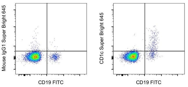
Invitrogen™ CD1c Monoclonal Antibody (L161), Super Bright™ 645, eBioscience™
Mouse Monoclonal Antibody
Supplier: Invitrogen™ 64001542


Description
Description: This L161 monoclonal antibody detects CD1c (also known as BDCA-1), a glycoprotein that is noncovalently linked to beta-2 microglobulin on thymocytes and antigen presenting cells such as dendritic and Langerhans cells. This molecule is also expressed on some circulating and marginal zone B cells, as well as in lymph nodes and germinal centers. CD1c is involved in the presentation of lipid antigens such as microbial fatty acids to effector T cells during the adaptive immune response. Finally, alternative splicing gives rise to three different isoforms of CD1c (soluble, membrane, and cytoplasmic/soluble isoforms). Applications Reported: This L161 antibody has been reported for use in flow cytometric analysis. Applications Tested: This L161 antibody has been pre-diluted and tested by flow cytometric analysis of normal human peripheral blood cells. This may be used at 5 μL (0.25 μg) per test. A test is defined as the amount (μg) of antibody that will stain a cell sample in a final volume of 100 μL. Cell number should be determined empirically but can range from 10^5 to 10^8 cells/test. Super Bright 645 is a tandem dye that can be excited with the violet laser line (405 nm) and emits at 645 nm. We recommend using a 660/20 bandpass filter. Please make sure that your instrument is capable of detecting this fluorochrome. When using two or more Super Bright dye-conjugated antibodies in a staining panel, it is recommended to use Super Bright Complete Staining...
This gene encodes a member of the CD1 family of transmembrane glycoproteins, which are structurally related to the major histocompatibility complex (MHC) proteins and form heterodimers with beta-2-microglobulin. The CD1 proteins mediate the presentation of primarily lipid and glycolipid antigens of self or microbial origin to T cells. The human genome contains five CD1 family genes organized in a cluster on chromosome 1. The CD1 family members are thought to differ in their cellular localization and specificity for particular lipid ligands. The protein encoded by this gene localizes to late endosomes and lysosomes via a tyrosine-based motif in the cytoplasmic tail, and requires vesicular acidification to bind lipid antigens.Specifications
| CD1c | |
| Monoclonal | |
| 5 μL/Test | |
| PBS with BSA and 0.09% sodium azide; pH 7.2 | |
| P29017 | |
| CD1C | |
| Affinity chromatography | |
| RUO | |
| 911 | |
| 4°C, store in dark, DO NOT FREEZE! | |
| Liquid |
| Flow Cytometry | |
| L161 | |
| Super Bright 645 | |
| CD1C | |
| BDCA1; canCD1c; CD1; CD1A; CD1C; CD1C antigen, c polypeptide; CD1c molecule; cortical thymocyte antigen CD1C; differentiation antigen CD1-alpha-3; R7; RP11-101J8.3; T-cell surface glycoprotein CD1c | |
| Mouse | |
| 100 Tests | |
| Primary | |
| Human | |
| Antibody | |
| IgG1 κ |
Product Content Correction
The Fisher Scientific Encompass Program offers items which are not part of our distribution portfolio. These products typically do not have pictures or detailed descriptions. However, we are committed to improving your shopping experience. Please use the form below to provide feedback related to the content on this product.
Product Title
Spot an opportunity for improvement?Share a Content Correction