Learn More
Invitrogen™ CD1d Monoclonal Antibody (51.1), PerCP-eFluor™ 710, eBioscience™, Invitrogen™


Description
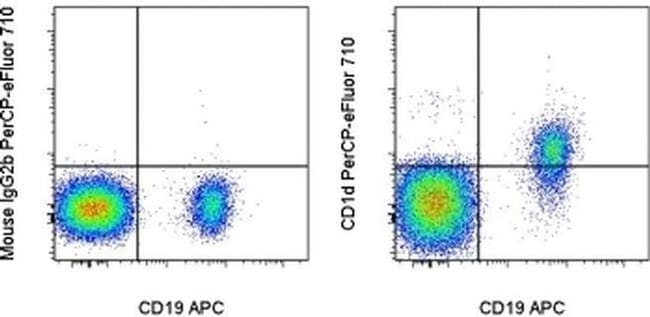
Description: The monoclonal antibody 51.1 reacts with human CD1d, a member of the CD1 family with similarity to the non-polymorphic MHC Class I-like molecules. CD1d is a highly conserved single transmembrane receptor of the Immunoglobulin Superfamily. CD1d can associate with beta-microglobulin another feature showing similarity to MHC class I molecules, but can also exist as a nonglycosylated protein not in association with beta microglobulin. This suggests different control mechanisms for presenting glycolipid containing molecules to CD1d reactive NKT cells. Expression of CD1d is found on B cells of the periphery, in resting monocytes and cortical thymocytes. On intestinal epithelial cells (IEC) expression is polarized. Expression can also be found at low levels intracellularly in hepatocytes. In HCV (hepatitis C virus) livers, CD1d is highly expressed compared to normal controls. The 51.1 monoclonal antibody has been shown to have functional activity; blocking the interaction of CD1d transfected cells with NKT cells. Applications Reported: This 51.1 antibody has been reported for use in flow cytometric analysis. Applications Tested: This 51.1 antibody has been pre-titrated and tested by flow cytometric analysis of normal human peripheral blood cells. This can be used at 5 μL (0.125 μg) per test. A test is defined as the amount (μg) of antibody that will stain a cell sample in a final volume of 100 μL.
Specifications
Specifications
| Antigen | CD1d |
| Applications | Flow Cytometry |
| Classification | Monoclonal |
| Clone | 51.1 |
| Concentration | 5 μL/Test |
| Conjugate | PerCP-eFluor 710 |
| Formulation | PBS with BSA and 0.09% sodium azide; pH 7.2 |
| Gene | CD1D |
| Gene Accession No. | P15813 |
| Gene Alias | AI747460; Antigen-presenting glycoprotein CD1d; antigen-presenting glycoprotein CD1d1; Cd1; CD1.1; CD1A; Cd1d; CD1D antigen; CD1D antigen, d polypeptide; CD1d molecule; CD1d.1; Cd1d1; CD1d1 antigen; CD1d1 molecule; differentiation antigen CD1-alpha-3; HMC class I antigen-like glycoprotein CD1D; Ly-38; R3; R3G1; T-cell surface glycoprotein CD1d; T-cell surface glycoprotein CD1d1; thymocyte antigen CD1D |
| Show More |
Frequently Asked Questions (FAQs)
Our options will depend on the samples you are analyzing.
If cell viability is not critical, you can store your stained samples at 4 degrees C or on ice overnight in the dark and analyze the following day.
For samples stained with eFluor organic fluorochromes, we recommend that cells be suspended in 100 uL of Flow Cytometry Staining Buffer (Cat. No. 00-4222) and 100 uL of eBioscience IC Fixation Buffer (Cat. No. 00-8222); samples can be incubated for up to 3 days at 4 degrees C in the dark. Alternatively, the 1-step Fix/Lyse Solution (Cat. No. 00-5333) can be used. This is a great option when working with whole blood but also works for other cell types.
Yes, the eFluor Organic fluorochromes can be used for intracellular staining. The eFluor organic fluorochromes maintain bright signal and require minimal changes in compensation when fixed with eBioscience IC Fixation Buffer (Cat. No. 00-8222-49) and Permeabilization Buffer (Cat. No. 00-8333-56) or 1-step Fix/Lyse Solution (Cat. No. 00-5333-54, 00-5333-57) (as compared to live cells).
The eFluor Organic fluorochromes and eVolve QDots can be used with flow staining buffers containing PBS and protein.
The eFluor Organic Dyes (eFluor 450, APC-eFluor 780, PerCP-eFluor 710, eFluor 710) are conventional fluorochromes. In contrast, the eVolve line of products are Quantum dots.
As with other fluorochromes, we recommend minimal exposure to light to maintain optimal signal.
For Research Use Only.
By clicking Submit, you acknowledge that you may be contacted by Fisher Scientific in regards to the feedback you have provided in this form. We will not share your information for any other purposes. All contact information provided shall also be maintained in accordance with our Privacy Policy.