Learn More
Invitrogen™ CD20 Monoclonal Antibody (QCH6A7), PerCP-eFluor™ 710, eBioscience™, Invitrogen™


Description
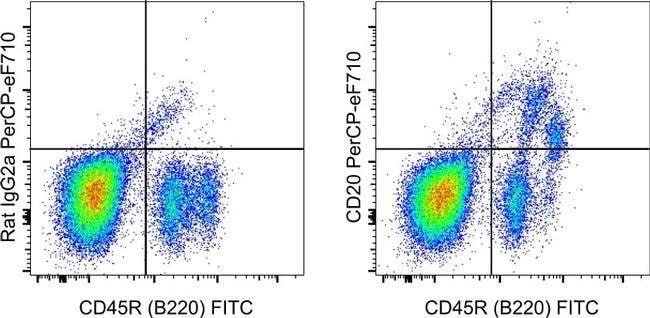
Description: The monoclonal antibody QCH6A7 reacts with mouse CD20. CD20 is a B cell specific, 32-kDa membrane protein that is expressed on late pro-B cells through memory B cells in the B cell lineage. CD20 is not found on hematopoietic stem cells, early pro-B cells or later mature plasma cells. CD20 is involved in intracellular calcium regulation and plays a role in B cell proliferation. CD20 is intially expressed after the induction of CD19 during development and is coexpressed with IgM during the pre-B to immature B cell transition in bone marrow. Expression of CD20 increases during maturation with almost all mature B cells expressing some level of CD20. Unlike other B cell antigens, CD20 is not shed or internalized upon antibody binding. Monoclonal antibodies against the CD20 antigen may be used to deplete B cells and CD20 has proven to be an effective immunotheraputic target in B cell lymphomas and several autoimmune diseases including rheumatoid arthritis and type I diabetes. CD20 consists of 4 transmembrane domains and a 44 amino acid extracellular domain which contains the QCH6A7 epitope. Staining with clone QCH6A7 provides significantly improved staining intensity compared to clone AISB12 in flow cytometric assays. Applications Reported: This QCH6A7 antibody has been reported for use in flow cytometric analysis. Applications Tested: This QCH6A7 antibody has been tested by flow cytometric analysis of mouse bone marrow cells and mouse splenocytes.
Specifications
Specifications
| Antigen | CD20 |
| Applications | Flow Cytometry |
| Classification | Monoclonal |
| Clone | QCH6A7 |
| Concentration | 0.2 mg/mL |
| Conjugate | PerCP-eFluor 710 |
| Formulation | PBS with 0.09% sodium azide; pH 7.2 |
| Gene | Ms4a1 |
| Gene Accession No. | P19437 |
| Gene Alias | AA960661; APY; ATOPY; B1; B-cell differentiation antigen Ly-44; B-lymphocyte antigen CD20; B-lymphocyte cell-surface antigen B1; B-lymphocyte surface antigen B1; Bp35; Cd20; CD20 antigen; CD20 cell surface protein; CD20 receptor; CVID5; EGK_06167; Fc epsilon receptor I beta chain; Fc Fragment of IgE high affinity I receptor for beta polypeptide; FCER1B; High affinity immunoglobulin epsilon receptor subunit beta; IgE Fc receptor subunit beta; IGEL; IGER; IGHER; LEU16; LEU-16; Leukocyte surface antigen Leu-16; Ly44; Ly-44; Lymphocyte antigen 44; membrane spanning 4-domains A1; membrane-spanning 4-domains subfamily A member 1; membrane-spanning 4-domains, subfamily A, member 1; membrane-spanning 4-domains, subfamily A, member 2; MGC3969; MS4A1; MS4A2; S7 |
| Show More |
Frequently Asked Questions (FAQs)
Our options will depend on the samples you are analyzing.
If cell viability is not critical, you can store your stained samples at 4 degrees C or on ice overnight in the dark and analyze the following day.
For samples stained with eFluor organic fluorochromes, we recommend that cells be suspended in 100 uL of Flow Cytometry Staining Buffer (Cat. No. 00-4222) and 100 uL of eBioscience IC Fixation Buffer (Cat. No. 00-8222); samples can be incubated for up to 3 days at 4 degrees C in the dark. Alternatively, the 1-step Fix/Lyse Solution (Cat. No. 00-5333) can be used. This is a great option when working with whole blood but also works for other cell types.
The eFluor Organic fluorochromes and eVolve QDots can be used with flow staining buffers containing PBS and protein.
Safety and Handling
By clicking Submit, you acknowledge that you may be contacted by Fisher Scientific in regards to the feedback you have provided in this form. We will not share your information for any other purposes. All contact information provided shall also be maintained in accordance with our Privacy Policy.