Learn More
Invitrogen™ CD27 Monoclonal Antibody (O323), Brilliant Ultra Violet™ 737, eBioscience™, Invitrogen™


Description
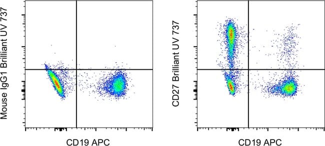
The O323 monoclonal antibody reacts with human CD27, a lymphocyte-specific member of the TNFR superfamily. CD27 is expressed by a subset of thymocytes and virtually all mature T cells and is upregulated upon T-cell stimulation. CD27 binds to CD70, and through this interaction, plays an important role in T cell-B cell interaction. Applications Reported: This O323 antibody has been reported for use in flow cytometric analysis. Applications Tested: This O323 antibody has been pre-diluted and tested by flow cytometric analysis of normal human peripheral blood cells. This may be used at 5 μL (0.25 μg) per test. A test is defined as the amount (μg) of antibody that will stain a cell sample in a final volume of 100 μL. Cell number should be determined empirically but can range from 10^5 to 10^8 cells/test. Brilliant Ultraviolet 737 is a tandem dye that emits at 732 nm and is intended for use on cytometers equipped with an ultraviolet (355 nm) laser.
Specifications
Specifications
| Antigen | CD27 |
| Applications | Flow Cytometry |
| Classification | Monoclonal |
| Clone | O323 |
| Concentration | 5 μL/Test |
| Conjugate | Brilliant Ultraviolet 737 |
| Formulation | PBS with BSA and 0.09% sodium azide; pH 7.2 |
| Gene | CD27 |
| Gene Accession No. | P26842 |
| Gene Alias | CD antigen 27; CD27; CD27 antigen; CD27 molecule; CD27L receptor; LPFS2; S152; S152. LPFS2; sCD27; soluble CD27; T cell activation antigen S152; T14; T-cell activation antigen CD27; TNFRSF7; TNFSF7; Tp55; Tumor necrosis factor receptor superfamily member 7; tumor necrosis factor receptor superfamily, member 7 |
| Show More |
Safety and Handling
By clicking Submit, you acknowledge that you may be contacted by Fisher Scientific in regards to the feedback you have provided in this form. We will not share your information for any other purposes. All contact information provided shall also be maintained in accordance with our Privacy Policy.