Promotional price valid on web orders only. Your contract pricing may differ. Interested in signing up for a dedicated account number?
Learn More

Invitrogen™ CD273 (B7-DC) Monoclonal Antibody (122), Super Bright™ 436, eBioscience™
GREENER_CHOICE

Catalog No. 62997282
Encompass_Preferred
Change view
Click to view available options
Quantity:
100 μg
1 product options available for selection
Product selection table with 1 available options. Use arrow keys to navigate and Enter or Space to select.
Catalog No. Quantity
62997282 100 μg
Use arrow keys to navigate between rows. Press Enter or Space to select a product option. 1 options available.
1 options
Catalog No. 62997282 Supplier Invitrogen™ Supplier No. 62997282
Add to Cart
Edge
Add to Cart

Rat Monoclonal Antibody

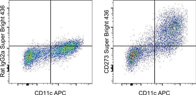
The 122 monoclonal antibody reacts with mouse B7-DC, also known as PD-L2. B7-DC, a member of the B7 family, has a predicted molecular weight of approximately 25 kDa and belongs to the Ig superfamily. The mouse B7-DC has a short cytoplasmic tail (4aa). B7-DC is primarily expressed by subpopulations of dendritic cells and monocytes/macrophages in the mouse. Although B7-DC has structural and sequence similarities to the B7 family, it does not bind CD28/CTLA-4, rather it is a ligand for PD-1. The interactions between PD-1 and B7-DC/PD-L2 have been reported to be involved in costimulation or suppression of T cell proliferation depending on state of cellular activation. 122 has been demonstrated to block binding of TY25 (Product # 14-5986), another mAb specific for mouse B7-DC. Applications Reported: This 122 antibody has been reported for use in flow cytometric analysis. Applications Tested: This 122 antibody has been tested by flow cytometric analysis of bone marrow derived dendritic cells. This may be used at less than or equal to 1.0 μg per test. A test is defined as the amount (μg) of antibody that will stain a cell sample in a final volume of 100 μL. Cell number should be determined empirically but can range from 10^5 to 10^8 cells/test. It is recommended that the antibody be carefully titrated for optimal performance in the assay of interest. Super Bright 436 can be excited with the violet laser line (405 nm) and emits at 436 nm. We recommend using a...

Programmed death-ligand 2 (PD-L2), or B7-DC, is a member of the B7 ligand family within the immunoglobulin superfamily that, along with programmed death-ligand 1 (PD-L1), acts as a ligand for programmed cell death protein 1 (PD-1). Though expressed primarily in dendritic cells, PD-L2 expression can be induced on a wide variety of immune and non-immune cells depending on the microenvironment. PD-L2 expression is particularly upregulated in the presence of Th2 cytokine, IL-4, as well as Th1 cytokines, TNF-alpha and IFN-gamma to a lesser degree. While generally expressed at lower levels compared to PD-L1, PD-L2 demonstrates a 2 to 6 times higher relative affinity to PD-1 than PD-L1. PD-1 and its ligands are referred to as inhibitory immune checkpoint molecules in that they provide useful negative feedback during physiological homeostasis. Ligation of PD-L2 or PD-L1 inhibits activation, proliferation, and cytokine secretion (e.g. IFN-gamma, IL-10) in T cells, ultimately dampening immune response. Conversely, studies have shown that PD-L2 can also stimulate T cell proliferation and cytokine production, even in PD-1-deficient T cells, suggesting additional receptors. Recent studies have concluded that PD-L2 also binds to a second receptor, repulsive guidance molecule b (RGMb), which was originally identified as a receptor for bone morphogenetic proteins (BMPs). RGMb is expressed in the central nervous system, as well as in macrophages, however, its role in immunity is only beginning to emerge. Interaction between PD-L2 and RGMb regulates the development of respiratory tolerance in the lung through BMP and/or neogenin signaling pathways. The naturally occurring human PD-L2 monomer consists of a 201-amino-acid extracellular domain, a 21-amino-acid transmembrane domain, and a 32-amino-acid cytoplasmic domain.
TRUSTED_SUSTAINABILITY

Specifications

Antigen CD273 (B7-DC)
Applications Flow Cytometry
Classification Monoclonal
Clone 122
Concentration 0.2 mg/mL
Conjugate Super Bright 436
Formulation PBS with BSA and 0.09% sodium azide; pH 7.2
Gene PDCD1LG2
Gene Accession No. Q9WUL5
Gene Alias B7 dendritic cell molecule; B7DC; B7-DC; bA574F11.2; Btdc; Butyrophilin B7-DC; butyrophilin-like protein; CD273; F730015O22Rik; MGC124039; MGC124040; PD1 ligand 2; PD-1 ligand 2; PD-1-ligand 2; PDCD1 ligand 2; PDCD1L2; Pdcd1lg2; Pdl2; PD-L2; Programmed cell death 1 ligand 2; programmed death ligand 2
Gene Symbols PDCD1LG2
Host Species Rat
Purification Method Affinity chromatography
Quantity 100 μg
Regulatory Status RUO
Primary or Secondary Primary
Gene ID (Entrez) 58205
Target Species Mouse
Content And Storage 4°C, store in dark, DO NOT FREEZE!
Product Type Antibody
Form Liquid
Isotype IgG2a κ
Show More Show Less
Can I use the OneComp and UltraComp eBeads microspheres with Super Bright-conjugated antibodies?

UltraComp eBeads microspheres (Cat. No. 01-2222) are recommended for use with Super Bright dyes.
Note: Super Bright Staining Buffer (Cat. No. SB-4400) is not compatible with UltraComp eBeads microspheres (Cat. No. 01-2222-41, 00-2222-42). If using UltraComp eBeads microspheres as a compensation tool, solely use Flow Cytometry Stain Buffer (Cat. No. 00-4222-26, 00-4222-57) for any antibody dilutions.

In some experiments, we have observed that compensation values for Super Bright 780- and Brilliant Violet 785- or Brilliant Violet 786-conjugated antibodies are higher in the violet 450/50 channel when using UltraComp eBeads microspheres as compared to single-color stained cells. In such circumstances, we would recommend setting compensation with cells. We have also observed this in some experiments using AbC Total Antibody Compensation beads, both with Super Bright 780 and Brilliant Violet 786. We have not tested Brilliant Violet 785 with the AbC beads.

Can I prepare an antibody cocktail containing Super Bright Staining Buffer and Super Bright-conjugated antibodies ahead of time?

We recommend that the antibody cocktails containing Super Bright-conjugated antibodies and Super Bright Staining Buffer are prepared fresh prior to staining. Discard any unused portions. We do not recommend overnight storage of prepared cocktails.

Can I fix my cells after staining with Super Bright-conjugated antibodies? How long can fixed cells be stored prior to analysis?

Samples that have been stained with antibodies conjugated to Super Bright dyes may be stored for up to three days, at 2-8°C, in the dark, using either IC Fixation Buffer (Cat. No. 00-8222) or 1-step Fix/Lyse Buffer (Cat. No. 00-5333) with no significant effect on brightness or compensation.

Are the Super Bright dyes sensitive to methanol fixation?

Super Bright dyes are stable in methanol-based fixation buffers.

Can Super Bright-conjugated antibodies be used in combination with intracellular (IC Fixation/Permeabilization) or intranuclear (Foxp3 fixation/permeabilization) staining?

Yes, Super Bright-conjugated antibodies are stable in formaldehyde-based fixation buffers and permeabilization buffers, such as the IC Fixation and Permeabilization Buffer Set (Cat. No. 88-8824) and the Foxp3/Transcription Factor Staining Buffer Set (Cat. No. 00-5523).

Which laser do I use to detect Super Bright-conjugated antibodies?

The violet laser (405 nm) should be used to excite Super Bright-conjugated antibodies.

Can the Super Bright Staining Buffer be used with other polymer dyes?

Yes, the Super Bright Staining Buffer (Cat. No. SB-4400) is compatible with other polymer dyes (i.e., Brilliant Violet dyes) and is useful for minimizing any non-specific polymer interactions when two or more of these dyes are used in combination.

Can Super Bright-conjugated antibodies be used in combination with other polymer dyes? What buffer should I use when using more than one Super Bright or polymer dye?

When using two or more Super Bright dyes, or when using Super Bright dyes in combination with other polymer dyes (i.e., Brilliant Violet dyes), we recommend using the Super Bright Staining Buffer (Cat. No. SB-4400) to minimize any non-specific polymer interactions between these fluorochromes. For specific instructions for use, please refer to the product technical data sheet.

Are there recommended tubes for staining with Super Bright-conjugated antibodies?

We recommend using polystyrene tubes (for example, FACS tubes) for staining with your Super Bright-conjugated antibodies. If using polypropylene (for example, Eppendorf tubes), protecting from light is critical.

Is there a specific buffer I should use when staining with Super Bright-conjugated antibodies?

No special buffer is required when using a single Super Bright-conjugated antibody in a panel.
When using more than one Super Bright dye, or when using Super Bright dyes in combination with other polymer dyes (i.e., Brilliant Violet dyes), we recommend using the Super Bright Staining Buffer (Cat. No. SB-4400) to minimize any non-specific polymer interactions between these fluorochromes. For specific instructions for use, please refer to the product technical data sheet.

How does the protocol for staining with Super Bright-conjugated antibodies differ from staining with conventional organic fluorochrome and eVolve conjugated antibodies?

Antibodies conjugated to Super Bright dyes can be used similarly to traditional fluorochromes. If multiple Super Bright dyes are used in combination with each other or in combination with other polymer dyes (i.e., Brilliant Violet dyes), then the use of Super Bright Staining Buffer (Cat. No. SB-4400) is recommended to minimize any non-specific polymer interactions between these fluorochromes. For specific instructions for use, please refer to the product technical data sheet.

Can the Super Bright dyes be used for non-flow applications?

Super Bright dyes have not been tested for applications other than flow cytometry.

What is the difference between the Super Bright and eVolve dyes?

Super Bright dyes are fluorochromes based on polymer technology and are excited by the violet laser. In contrast, eVolve dyes are based on Qdot nanocrystal technology and are maximally excited by the UV laser, although they may also be excited by violet, blue, yellow-green, and, depending on the eVolve, the red laser lines. The general shape of the emission spectra for Super Bright dyes will be more similar to traditional fluorochromes, while eVolve dyes have extremely narrow emission spectra. Please contact Tech Support (techsupport@thermofisher.com) for more information.

What are the Super Bright dyes?

eBioscience Super Bright dyes are a series of patent-pending fluorochromes that are based upon a fluorescent polymer and its tandems. Super Bright dyes have been developed for use in flow cytometry and can be excited by the violet laser (405 nm). The peak emission of each Super Bright dye is indicated by the number in the product description, e.g., "Super Bright 600" has a peak emission of 600 nm.

Are the Super Bright Dyes photo-labile?

As with other fluorochromes, we recommend minimal exposure to light to maintain optimal signal.


Product Title
Select an issue

By clicking Submit, you acknowledge that you may be contacted by Fisher Scientific in regards to the feedback you have provided in this form. We will not share your information for any other purposes. All contact information provided shall also be maintained in accordance with our Privacy Policy.