Learn More
Invitrogen™ CD3 Monoclonal Antibody (OKT3), Super Bright™ 600, eBioscience™, Invitrogen™

Description
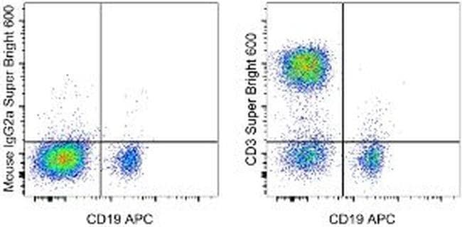
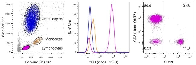
Description: The OKT3 monoclonal antibody reacts with an epitope on the epsilon-subunit within the human CD3 complex. The OKT3 antibody has been reported to have potent immunosuppressive properties in vivo and has been proven effective in the treatment of renal, heart and liver allograft rejection. The CD3 subunits, gamma, delta, and epsilon chains, are required for proper assembly, trafficking and surface expression of the TCR complex. CD3 is expressed by thymocytes in a developmentally regulated manner and by all mature T cells. Crosslinking of TCR initiates an intracellular biochemical pathway resulting in cellular activation and proliferation. Antibody clones OKT3 and SK7 see different epitopes. Applications Reported: This OKT3 antibody has been reported for use in flow cytometric analysis. Applications Tested: This OKT3 antibody has been pre-titrated and tested by flow cytometric analysis of normal human peripheral blood cells. This can be used at 5 μL (0.25 μg) per test. A test is defined as the amount (μg) of antibody that will stain a cell sample in a final volume of 100 μL. Cell number should be determined empirically but can range from 10^5 to 10^8 cells/test. Super Bright 600 is a tandem dye that can be excited with the violet laser line (405 nm) and emits at 600 nm. We recommend using a 610/20 bandpass filter. Please make sure that your instrument is capable of detecting this fluorochrome.
Specifications
Specifications
| Antigen | CD3 |
| Applications | Flow Cytometry |
| Classification | Monoclonal |
| Clone | OKT3 |
| Concentration | 5 μL/Test |
| Conjugate | Super Bright 600 |
| Formulation | PBS with BSA and 0.09% sodium azide; pH 7.2 |
| Gene | Cd3d |
| Gene Accession No. | P04234, P07766, P09693, P20963 |
| Gene Alias | 4930549J05Rik; A430104F18Rik; AI504783; antigen CD3D, delta polypeptide (TiT3 complex); antigen CD3E, epsilon polypeptide (TiT3 complex); antigen CD3G, gamma polypeptide; antigen CD3Z, zeta polypeptide; AW552088; Cd247; CD247 antigen; CD247 antigen, zeta subunit; Cd247 molecule; CD3; CD3 antigen delta chain; CD3 antigen delta polypeptide; CD3 antigen gamma chain; CD3 antigen, delta polypeptide; CD3 antigen, delta subunit; CD3 antigen, epsilon polypeptide; CD3 antigen, gamma polypeptide; CD3 antigen, zeta polypeptide; CD3 delta; CD3 epsilon; CD3 epsilon chain; CD3 epsilon subunit; CD3 epsilon subunit precursor; CD3 gamma-chain; CD3 glycoprotein; CD3 glycoprotein precursor; CD3 molecule delta polypeptide; CD3 molecule, delta; CD3 molecule, epsilon; CD3 molecule, epsilon polypeptide; CD3 molecule, gamma; CD3 molecule, gamma polypeptide; CD3 protein; CD3 TCR complex; CD3 type I transmembrane glycoprotein; CD3 type I transmembrane glycoprotein precursor; CD3 zeta chain; Cd3d; CD3D antigen delta; CD3D antigen, delta polypeptide (TiT3 complex); CD3d molecule; CD3d molecule, delta (CD3-TCR complex); CD3-DELTA; Cd3e; CD3E antigen, epsilon polypeptide; CD3E antigen, epsilon polypeptide (TiT3 complex); CD3e molecule; CD3e molecule, epsilon (CD3-TCR complex); CD3epsilon; CD3-epsilon; Cd3-eta; Cd3g; CD3G antigen, gamma polypeptide; CD3g antigen, gamma polypeptide (TiT3 complex); CD3g molecule; CD3g molecule, epsilon (CD3-TCR complex); CD3g molecule, gamma (CD3-TCR complex); CD3-GAMMA; Cd3h; CD3Q; Cd3z; CD3Z antigen, zeta polypeptide (TiT3 complex); Cd3zeta; Cd3-zeta; CD3zeta chain; CD3-zeta/eta; Ctg3; Ctg-3; FLJ18683; IMD17; IMD18; IMD19; IMD25; Leu-4; OKT3, delta chain; T cell antigen receptor complex epsilon subunit of T3; T3/TCR complex; T3d; T3e; T3g; T3Z; T-cell antigen receptor complex, epsilon subunit of T3; T-cell antigen receptor complex, gamma subunit of T3; T-cell antigen receptor complex, zeta subunit of CD3; T-cell receptor CD3 epsilon chain; T-cell receptor CD3 epsilon subunit; T-cell receptor CD3 subunit zeta; T-cell receptor CD3, subunit zeta; T-cell receptor T3 delta chain; T-cell receptor T3 eta chain; T-cell receptor T3 gamma chain; T-cell receptor T3 zeta chain; T-cell receptor zeta chain; T-cell surface antigen T3/Leu-4 epsilon chain; T-cell surface glycoprotein CD3 delta chain; T-cell surface glycoprotein CD3 epsilon chain; T-cell surface glycoprotein CD3 gamma chain; T-cell surface glycoprotein CD3 zeta chain; T-cell surface protein; TcR CD3 delta-chain; TcR CD3 gamma-chain; TCR zeta chain; TCR zeta chain subunit; TCRE; Tcrk; TCRZ; TCRzeta; TiT3 complex; type I transmembrane protein; T-cell surface molecule CD3 |
| Show More |
Frequently Asked Questions (FAQs)
UltraComp eBeads microspheres (Cat. No. 01-2222) are recommended for use with Super Bright dyes.
Note: Super Bright Staining Buffer (Cat. No. SB-4400) is not compatible with UltraComp eBeads microspheres (Cat. No. 01-2222-41, 00-2222-42). If using UltraComp eBeads microspheres as a compensation tool, solely use Flow Cytometry Stain Buffer (Cat. No. 00-4222-26, 00-4222-57) for any antibody dilutions.
In some experiments, we have observed that compensation values for Super Bright 780- and Brilliant Violet 785- or Brilliant Violet 786-conjugated antibodies are higher in the violet 450/50 channel when using UltraComp eBeads microspheres as compared to single-color stained cells. In such circumstances, we would recommend setting compensation with cells. We have also observed this in some experiments using AbC Total Antibody Compensation beads, both with Super Bright 780 and Brilliant Violet 786. We have not tested Brilliant Violet 785 with the AbC beads.
We recommend that the antibody cocktails containing Super Bright-conjugated antibodies and Super Bright Staining Buffer are prepared fresh prior to staining. Discard any unused portions. We do not recommend overnight storage of prepared cocktails.
Samples that have been stained with antibodies conjugated to Super Bright dyes may be stored for up to three days, at 2-8°C, in the dark, using either IC Fixation Buffer (Cat. No. 00-8222) or 1-step Fix/Lyse Buffer (Cat. No. 00-5333) with no significant effect on brightness or compensation.
Super Bright dyes are stable in methanol-based fixation buffers.
Yes, Super Bright-conjugated antibodies are stable in formaldehyde-based fixation buffers and permeabilization buffers, such as the IC Fixation and Permeabilization Buffer Set (Cat. No. 88-8824) and the Foxp3/Transcription Factor Staining Buffer Set (Cat. No. 00-5523).
For Research Use Only.
By clicking Submit, you acknowledge that you may be contacted by Fisher Scientific in regards to the feedback you have provided in this form. We will not share your information for any other purposes. All contact information provided shall also be maintained in accordance with our Privacy Policy.