Learn More
Invitrogen™ CD304 (Neuropilin-1) Monoclonal Antibody (3DS304M), PerCP-eFluor™ 710, eBioscience™, Invitrogen™


Description
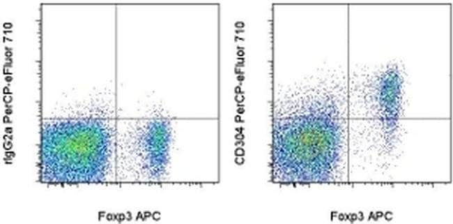
Description: The monoclonal antibody 3DS304M recognizes mouse CD304 (Neuropilin-1), a type 1 transmembrane glycoprotein present on the surface of multiple cell types, including: regulatory T cells (Treg), NKT cells, DC, certain types of stem cells, neurons, endothelial cells, and some neoplastic cells. Neuropilin-1, in complex with plexins, serves as a co-receptor for type 3 semaphorins in growing neurons. It is also involved in the process of angiogenesis being a part of a functional receptor for VEGF in endothelial cells. In mice, Neuropilin-1 is expressed on thymus-derived natural Treg but not peripherally induced Treg. In addition, transient expression on recently activated non-regulatory T cells has been observed. Neuropilin-1 has been used as a marker of recent thymic emigrants in the mouse iNKT cell population. It has been shown that Neuropilin-1 forms a complex with TGF beta receptors, activating the latent form of TGF beta (LAP-TGF beta 1) and augmenting canonical Smad2/3 signaling. This 3DS304M antibody will recognize formaldehyde-fixed as well as methanol-treated epitopes. Applications Reported: This 3DS304M antibody has been reported for use in flow cytometric analysis. Applications Tested: This 3DS304M antibody has been tested by flow cytometric analysis of mouse splenocytes. This can be used at less than or equal to 0.06 μg per test.
Specifications
Specifications
| Antigen | CD304 (Neuropilin-1) |
| Applications | Flow Cytometry |
| Classification | Monoclonal |
| Clone | 3DS304M |
| Concentration | 0.2 mg/mL |
| Conjugate | PerCP-eFluor 710 |
| Formulation | PBS with 0.09% sodium azide; pH 7.2 |
| Gene | NRP1 |
| Gene Accession No. | P97333 |
| Gene Alias | A5 protein; BDCA4; C530029I03; CD304; Neuropilin; neuropilin 1; Neuropilin1; neuropilin-1; Neuropilin-1 precursor (A5 protein); NP1; NP-1; Npn1; NPN-1; NRP; NRP 1; Nrp1; sNRP 1; sNRP1; soluble NRP 1; soluble NRP1; transmembrane receptor; vascular endothelial cell growth factor 165 receptor; VEGF165R |
| Show More |
Frequently Asked Questions (FAQs)
Our options will depend on the samples you are analyzing.
If cell viability is not critical, you can store your stained samples at 4 degrees C or on ice overnight in the dark and analyze the following day.
For samples stained with eFluor organic fluorochromes, we recommend that cells be suspended in 100 uL of Flow Cytometry Staining Buffer (Cat. No. 00-4222) and 100 uL of eBioscience IC Fixation Buffer (Cat. No. 00-8222); samples can be incubated for up to 3 days at 4 degrees C in the dark. Alternatively, the 1-step Fix/Lyse Solution (Cat. No. 00-5333) can be used. This is a great option when working with whole blood but also works for other cell types.
Yes, the eFluor Organic fluorochromes can be used for intracellular staining. The eFluor organic fluorochromes maintain bright signal and require minimal changes in compensation when fixed with eBioscience IC Fixation Buffer (Cat. No. 00-8222-49) and Permeabilization Buffer (Cat. No. 00-8333-56) or 1-step Fix/Lyse Solution (Cat. No. 00-5333-54, 00-5333-57) (as compared to live cells).
The eFluor Organic Dyes (eFluor 450, APC-eFluor 780, PerCP-eFluor 710, eFluor 710) are conventional fluorochromes. In contrast, the eVolve line of products are Quantum dots.
As with other fluorochromes, we recommend minimal exposure to light to maintain optimal signal.
For Research Use Only.
By clicking Submit, you acknowledge that you may be contacted by Fisher Scientific in regards to the feedback you have provided in this form. We will not share your information for any other purposes. All contact information provided shall also be maintained in accordance with our Privacy Policy.