Promotional price valid on web orders only. Your contract pricing may differ. Interested in signing up for a dedicated account number?
Learn More
Learn More
CD326 (EpCAM) Monoclonal Antibody (1B7), Super Bright™ 645, eBioscience™, Invitrogen™

Mouse Monoclonal Antibody
$432.00
Specifications
| Antigen | CD326 (EpCAM) |
|---|---|
| Clone | 1B7 |
| Applications | Flow Cytometry |
| Classification | Monoclonal |
| Conjugate | Super Bright 645 |
| Catalog Number | Mfr. No. | Quantity | Price | Quantity & Availability | |||||
|---|---|---|---|---|---|---|---|---|---|
| Catalog Number | Mfr. No. | Quantity | Price | Quantity & Availability | |||||
64-932-642

|
Invitrogen™
64932642 |
100 Tests |
Each of 1 for $432.00
|
|
|||||
Description
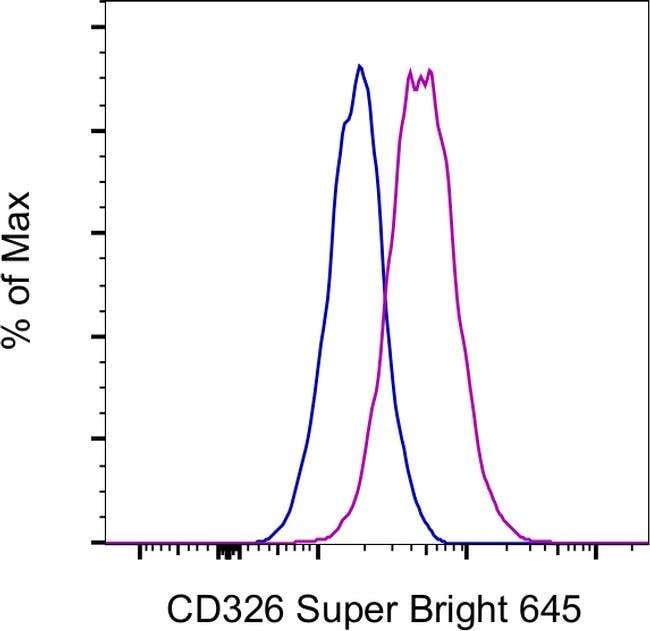
Description: EpCAM (Epithelial cell adhesion molecule, CD326, KSA, TROP1) is a 40 kD cell-surface adhesion molecule participating in homophilic, calcium-independent cell-cell interactions. EpCAM is a type-I transmembrane protein, and is expressed primarily on the basolateral surface of most epithelia. Although normal epithelia express low levels of EpCAM, increased expression has been correlated with increased proliferation and progression to a mesenchymal phenotype. EpCAM has also been used as a diagnostic marker on circulating metastatic carcinoma cells, while cancer cells of non-epithelial origin do not express EpCAM. Applications Reported: This 1B7 antibody has been reported for use in flow cytometric analysis. Applications Tested: This 1B7 antibody has been pre-diluted and tested by flow cytometric analysis of A549 cells. This may be used at 5 μL (0.5 μg) per test. A test is defined as the amount (μg) of antibody that will stain a cell sample in a final volume of 100 μL. Cell number should be determined empirically but can range from 10^5 to 10^8 cells/test. Super Bright 645 is a tandem dye that can be excited with the violet laser line (405 nm) and emits at 645 nm. We recommend using a 660/20 bandpass filter. Please make sure that your instrument is capable of detecting this fluorochrome. When using two or more Super Bright dye-conjugated antibodies in a staining panel, it is recommended to use Super Bright Complete Staining Buffer (Product No.
Ep-CAM (epithelial adhesion molecule, epithelial specific antigen, ESA) is a transmembrane glycoprotein expressed in the epithelium with a molecular weight of approximately 40 kDa, which functions as an epithelial cell adhesion molecule. Ep-CAM functions as a homotypic calcium-independent cell adhesion molecule, and has a direct impact on cell cycle, proliferation and metabolism of epithelial cells and fibroblasts due to its ability to rapidly induce the proto-oncogene c-myc and the cell cycle regulating genes cyclin A and E. Ep-CAM mediates Ca2+-independent homotypic interactions. Formation of Ep-CAM-mediated adhesions have a negative regulatory effect on adhesions mediated by classic cadherins, which may have strong effects on the differentiation and growth of epithelial cells. Ep-CAM overexpression was suggested to be associated with enhanced epithelial proliferation. Ep-CAM is highly expressed in human carcinomas, and is a marker for tumors of epithelial lineage. Ep-CAM is expressed on baso-lateral cell surface in most simple epithelia and many carcinoma types. Also, Ep-CAM reportedly distinguishes adenocarcinomas from pleural mesotheliomas.Specifications
| CD326 (EpCAM) | |
| Flow Cytometry | |
| Super Bright 645 | |
| Mouse | |
| Human | |
| 4072 | |
| IgG1 κ | |
| 4°C, store in dark, DO NOT FREEZE! | |
| EPCAM |
| 1B7 | |
| Monoclonal | |
| Liquid | |
| RUO | |
| P16422 | |
| EPCAM | |
| Primary | |
| Antibody |
Spot an opportunity for improvement?Share a Content Correction
Product Content Correction
The Fisher Scientific Encompass Program offers items which are not part of our distribution portfolio. These products typically do not have pictures or detailed descriptions. However, we are committed to improving your shopping experience. Please use the form below to provide feedback related to the content on this product.
Product Title