Promotional price valid on web orders only. Your contract pricing may differ. Interested in signing up for a dedicated account number?
Learn More

Invitrogen™ CD366 (TIM3) Monoclonal Antibody (8B.2C12), Super Bright™ 600, eBioscience™
GREENER_CHOICE

Catalog No. 63587182
Encompass_Preferred
Change view
Click to view available options
Quantity:
100 μg
1 product options available for selection
Product selection table with 1 available options. Use arrow keys to navigate and Enter or Space to select.
Catalog No. Quantity
63-587-182 100 μg
Use arrow keys to navigate between rows. Press Enter or Space to select a product option. 1 options available.
1 options
Catalog No. 63-587-182 Supplier Invitrogen™ Supplier No. 63587182
Add to Cart
Edge
Add to Cart

Rat Monoclonal Antibody

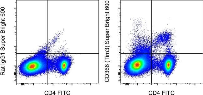
The 8B.2C12 monoclonal antibody reacts with mouse CD366 (TIM3), a Th1-specific cell surface protein. CD366 is a type I transmembrane protein and contains an immunoglobulin and a mucin-like domain in its extracellular portion and a tyrosine phosphorylation motif in its cytoplasmic portion. CD366 is expressed selectively by differentiated CD4+ Th1 and CD8+ Tc1, but is absent on Th2 and Tc2. Other hematopoietic cell types, including naive T cells, B cells, macrophages and dendritic cells, do not express CD366, at least at the protein level. Expression of CD366 is upregulated at a late stage of T cell differentiation on Th1 cells after 3 rounds of in vitro polarization suggesting a role for this molecule in the transport or effector function of Th1 cells rather than a contribution to T cell differentiation. In an experimental autoimmune encephalomyelitis (EAE) model, CD366 was shown to be expressed on most CD4+ and CD8+ T cells in the central nervous system at the onset of clinical signs of disease, while less than 2% of CD4+ cells in the periphery expressed CD366 after immunization. In this model, in vivo administration of 8B.2C12 resulted in a hyperacute and atypical disease phenotype. It is postulated that the engagement of CD366 during T cell activation results in the expansion and activation of macrophages and increased severity of autoimmune disease. The Tim gene family may have an important role in the regulation of autoimmunity and allergies. The 8B.2C12 a...

TIM3 (Hepatitis A virus cellular receptor 2, HAVCR2, T-cell immunoglobulin, mucin-dmain containing-3) is a 281 amino acid long, Type-1 Th1- specific cell surface glycoprotein expressed on terminally differentiated CD4+Th1 and CD8+Tc1 cells. TIM3 consists of an IgV-like domain, a mucin-like domain in the extracellular region, and a conserved Tyrosine phosphorylation motif in the cytoplasmic region. TIM3 is involved in macrophage activation and induction of autoimmune diseases. Further, TIM3 down-regulates aggressive Th1-mediated immune responses and facilitates in the development of immune tolerance. Pathological significance of TIM3 has been attributed to Experimental autoimmune encephalomyelitis (EAE), a Th-1 dependent autoimmune disease, and also enhances the severity of experimental autoimmune encephalomyelitis in mice.
TRUSTED_SUSTAINABILITY

Specifications

Antigen CD366 (TIM3)
Applications Flow Cytometry
Classification Monoclonal
Clone 8B.2C12
Concentration 0.2 mg/mL
Conjugate Super Bright 600
Formulation PBS with BSA and 0.09% sodium azide; pH 7.2
Gene Havcr2
Gene Accession No. Q8VIM0
Gene Alias CD366; FLJ14428; Havcr2; HAVcr-2; Hepatitis A virus cellular receptor 2; hepatitis A virus cellular receptor 2 homolog; kidney injury molecule-3; KIM-3; sCD366; soluble CD366; soluble TIM 3; T cell immunoglobulin mucin 3; T cell immunoglobulin mucin-3; T-cell immunoglobulin and mucin domain containing 3; T-cell immunoglobulin and mucin domain-containing protein 3; T-cell immunoglobulin mucin family member 3; T-cell immunoglobulin mucin receptor 3; T-cell membrane protein 3; Tim3; TIM-3; TIMD3; TIMD-3
Gene Symbols Havcr2
Host Species Rat
Purification Method Affinity chromatography
Quantity 100 μg
Regulatory Status RUO
Primary or Secondary Primary
Gene ID (Entrez) 171285
Target Species Mouse
Content And Storage 4°C, store in dark, DO NOT FREEZE!
Product Type Antibody
Form Liquid
Isotype IgG1 κ
Show More Show Less
Can I use the OneComp and UltraComp eBeads microspheres with Super Bright-conjugated antibodies?

UltraComp eBeads microspheres (Cat. No. 01-2222) are recommended for use with Super Bright dyes.
Note: Super Bright Staining Buffer (Cat. No. SB-4400) is not compatible with UltraComp eBeads microspheres (Cat. No. 01-2222-41, 00-2222-42). If using UltraComp eBeads microspheres as a compensation tool, solely use Flow Cytometry Stain Buffer (Cat. No. 00-4222-26, 00-4222-57) for any antibody dilutions.

In some experiments, we have observed that compensation values for Super Bright 780- and Brilliant Violet 785- or Brilliant Violet 786-conjugated antibodies are higher in the violet 450/50 channel when using UltraComp eBeads microspheres as compared to single-color stained cells. In such circumstances, we would recommend setting compensation with cells. We have also observed this in some experiments using AbC Total Antibody Compensation beads, both with Super Bright 780 and Brilliant Violet 786. We have not tested Brilliant Violet 785 with the AbC beads.

Can I prepare an antibody cocktail containing Super Bright Staining Buffer and Super Bright-conjugated antibodies ahead of time?

We recommend that the antibody cocktails containing Super Bright-conjugated antibodies and Super Bright Staining Buffer are prepared fresh prior to staining. Discard any unused portions. We do not recommend overnight storage of prepared cocktails.

Can I fix my cells after staining with Super Bright-conjugated antibodies? How long can fixed cells be stored prior to analysis?

Samples that have been stained with antibodies conjugated to Super Bright dyes may be stored for up to three days, at 2-8°C, in the dark, using either IC Fixation Buffer (Cat. No. 00-8222) or 1-step Fix/Lyse Buffer (Cat. No. 00-5333) with no significant effect on brightness or compensation.

Are the Super Bright dyes sensitive to methanol fixation?

Super Bright dyes are stable in methanol-based fixation buffers.

Can Super Bright-conjugated antibodies be used in combination with intracellular (IC Fixation/Permeabilization) or intranuclear (Foxp3 fixation/permeabilization) staining?

Yes, Super Bright-conjugated antibodies are stable in formaldehyde-based fixation buffers and permeabilization buffers, such as the IC Fixation and Permeabilization Buffer Set (Cat. No. 88-8824) and the Foxp3/Transcription Factor Staining Buffer Set (Cat. No. 00-5523).

Which laser do I use to detect Super Bright-conjugated antibodies?

The violet laser (405 nm) should be used to excite Super Bright-conjugated antibodies.

Can the Super Bright Staining Buffer be used with other polymer dyes?

Yes, the Super Bright Staining Buffer (Cat. No. SB-4400) is compatible with other polymer dyes (i.e., Brilliant Violet dyes) and is useful for minimizing any non-specific polymer interactions when two or more of these dyes are used in combination.

Can Super Bright-conjugated antibodies be used in combination with other polymer dyes? What buffer should I use when using more than one Super Bright or polymer dye?

When using two or more Super Bright dyes, or when using Super Bright dyes in combination with other polymer dyes (i.e., Brilliant Violet dyes), we recommend using the Super Bright Staining Buffer (Cat. No. SB-4400) to minimize any non-specific polymer interactions between these fluorochromes. For specific instructions for use, please refer to the product technical data sheet.

Are there recommended tubes for staining with Super Bright-conjugated antibodies?

We recommend using polystyrene tubes (for example, FACS tubes) for staining with your Super Bright-conjugated antibodies. If using polypropylene (for example, Eppendorf tubes), protecting from light is critical.

Is there a specific buffer I should use when staining with Super Bright-conjugated antibodies?

No special buffer is required when using a single Super Bright-conjugated antibody in a panel.
When using more than one Super Bright dye, or when using Super Bright dyes in combination with other polymer dyes (i.e., Brilliant Violet dyes), we recommend using the Super Bright Staining Buffer (Cat. No. SB-4400) to minimize any non-specific polymer interactions between these fluorochromes. For specific instructions for use, please refer to the product technical data sheet.

How does the protocol for staining with Super Bright-conjugated antibodies differ from staining with conventional organic fluorochrome and eVolve conjugated antibodies?

Antibodies conjugated to Super Bright dyes can be used similarly to traditional fluorochromes. If multiple Super Bright dyes are used in combination with each other or in combination with other polymer dyes (i.e., Brilliant Violet dyes), then the use of Super Bright Staining Buffer (Cat. No. SB-4400) is recommended to minimize any non-specific polymer interactions between these fluorochromes. For specific instructions for use, please refer to the product technical data sheet.

Can the Super Bright dyes be used for non-flow applications?

Super Bright dyes have not been tested for applications other than flow cytometry.

What is the difference between the Super Bright and eVolve dyes?

Super Bright dyes are fluorochromes based on polymer technology and are excited by the violet laser. In contrast, eVolve dyes are based on Qdot nanocrystal technology and are maximally excited by the UV laser, although they may also be excited by violet, blue, yellow-green, and, depending on the eVolve, the red laser lines. The general shape of the emission spectra for Super Bright dyes will be more similar to traditional fluorochromes, while eVolve dyes have extremely narrow emission spectra. Please contact Tech Support (techsupport@thermofisher.com) for more information.

What are the Super Bright dyes?

eBioscience Super Bright dyes are a series of patent-pending fluorochromes that are based upon a fluorescent polymer and its tandems. Super Bright dyes have been developed for use in flow cytometry and can be excited by the violet laser (405 nm). The peak emission of each Super Bright dye is indicated by the number in the product description, e.g., "Super Bright 600" has a peak emission of 600 nm.

Are the Super Bright Dyes photo-labile?

As with other fluorochromes, we recommend minimal exposure to light to maintain optimal signal.


Product Title
Select an issue

By clicking Submit, you acknowledge that you may be contacted by Fisher Scientific in regards to the feedback you have provided in this form. We will not share your information for any other purposes. All contact information provided shall also be maintained in accordance with our Privacy Policy.