Learn More
Invitrogen™ CD4 Monoclonal Antibody (N1UG0), eFluor™ 570, eBioscience™


Description
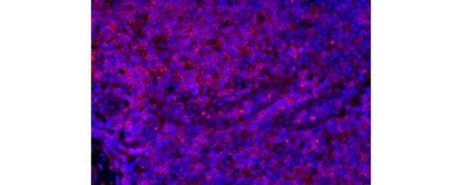
Description: The N1UG0 monoclonal antibody reacts with human CD4, a 59 kDa cell surface glycoprotein expressed by the majority of thymocytes, a subpopulation of mature T cells (T-helper cells) and in low levels on monocytes. CD4 is a receptor for the human immunodeficiency virus (HIV). The N1UG0 antibody is recommended for use in staining human formalin-fixed paraffin embedded tissue sections. Applications Reported: This N1UG0 antibody has been reported for use in immunohistochemical staining of formalin-fixed paraffin embedded tissue sections. Applications Tested: This N1UG0 antibody has been tested by immunohistochemistry of formalin-fixed paraffin embedded human tissue using low or high pH antigen retrieval at less than or equal to 20 μg/mL. It is recommended that the antibody be titrated for opitmal performance in the assay of interest. Filter Recommendation: When using this eFluor® 570 antibody conjugate, we recommend a filter that will capture the 570 emission wavelength (for example, Excitation 545/25, 565LP, Emission 605/70). A standard Alexa Fluor® 555 or TRITC filter is acceptable. Excitation: 555 nm; Emission: 570 nm.
Specifications
Specifications
| Antigen | CD4 |
| Applications | Immunohistochemistry (Paraffin), Immunocytochemistry |
| Classification | Monoclonal |
| Clone | N1UG0 |
| Concentration | 0.2 mg/mL |
| Conjugate | eFluor 570 |
| Formulation | PBS with 0.09% sodium azide; pH 7.2 |
| Gene | CD4 |
| Gene Accession No. | P01730 |
| Gene Alias | Activation B7-1 antigen; B7; B7.1; B7-1; BB1; B-lymphocyte activation antigen B7; CD28LG; CD28LG1; CD4; CD4 antigen; CD4 antigen (p55); CD4 antigen p55; Cd4 molecule; CD4 precursor; CD4 receptor; CD4, allele 1; cd4a; CD4mut; CD80; CD80 antigen (CD28 antigen ligand 1, B7-1 antigen); CD80 molecule; cell surface glycoprotein CD4; costimulatory factor CD80; costimulatory molecule variant IgV-CD80; CTLA-4 counter-receptor B7.1; fCD4; L3T4; LAB7; Leu-3; Ly-4; lymphocyte antigen CD4; lymphocyte antigen CD4 precursor; membrane protein; p55; T-cell differentiation antigen L3T4; T-cell surface antigen T4/Leu-3; T-cell surface glycoprotein CD4; T-cell surface glycoprotein CD4 precursor (T-cell surface antigen T4/Leu-3) (T-cell differentiation antigen L3T4); T-lymphocyte activation antigen CD80; W3/25; W3/25 antigen |
| Show More |
Frequently Asked Questions (FAQs)
Yes, the eFluor Organic fluorochromes can be used for intracellular staining. The eFluor organic fluorochromes maintain bright signal and require minimal changes in compensation when fixed with eBioscience IC Fixation Buffer (Cat. No. 00-8222-49) and Permeabilization Buffer (Cat. No. 00-8333-56) or 1-step Fix/Lyse Solution (Cat. No. 00-5333-54, 00-5333-57) (as compared to live cells).
The eFluor Organic fluorochromes and eVolve QDots can be used with flow staining buffers containing PBS and protein.
The eFluor Organic Dyes (eFluor 450, APC-eFluor 780, PerCP-eFluor 710, eFluor 710) are conventional fluorochromes. In contrast, the eVolve line of products are Quantum dots.
As with other fluorochromes, we recommend minimal exposure to light to maintain optimal signal.
By clicking Submit, you acknowledge that you may be contacted by Fisher Scientific in regards to the feedback you have provided in this form. We will not share your information for any other purposes. All contact information provided shall also be maintained in accordance with our Privacy Policy.