Learn More
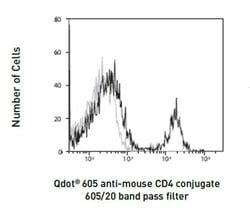
Invitrogen™ CD4 Monoclonal Antibody (RM4-5), Qdot™ 605
Rat Monoclonal Antibody
Supplier: Invitrogen™ Q10092

Description
Qdot™ Antibody (Ab) conjugates possess a bright fluorescence emission that makes them well suited for the detection of low-abundance extracellular proteins. Approximately the same size as R-phycoerythrin (R-PE) and compatible with existing organic fluorophore conjugates, Qdot™ Ab conjugates can be excited with any wavelength below their emission maximum, but are best excited by UV or violet light. The narrow, symmetric emission profiles of Qdot Ab conjugates allow for minimal compensation when using a single excitation source, and the very long stoke shifts enable better, more efficient multicolor assays using the 405 nm violet laser. Available in multiple colors for use in flow cytometry, these advantages make Qdot Ab conjugates powerful tools for antibody labeling and staining. Staining: Stain cells in any standard staining buffer, such as phosphate buffered saline (PBS) with 1% bovine serum albumin (BSA). We recommend analysis of cells within 18 hours of staining. If dilute reagent is used, dilute only the quantity of reagent to be used within one day. Qdot Ab conjugates may be mixed with other antibodies, but use the diluted conjugates on the day of dilution. Qdot Ab conjugates can be used for surface staining applications with most conventional sample preparation reagents, such as Cal-Lyse™ Lysing Solution and FIX & PERM™ reagents, with minimal effect on fluorescence. We have observed some batches of BD FACS™ Lysing Solution to interf...
The CD4 antigen is involved in the recognition of MHC class II molecules and is a co-receptor for HIV. CD4 is primarily expressed in a subset of T-lymphocytes, also referred to as T helper cells, but may also be expressed by other cells in the immune system, such as monocytes, macrophages, and dendritic cells. At the tissue level, CD4 expression may be detected in thymus, lymph nodes, tonsils, and spleen, and also in specific regions of the brain, gut, and other non-lymphoid tissues. CD4 functions to initiate or augment the early phase of T-cell activation through its association with the T-cell receptor complex and protein tyrosine kinase, Lck. It may also function as an important mediator of direct neuronal damage in infectious and immune-mediated diseases of the central nervous system. Multiple alternatively spliced transcripts have been identified in this gene [RefSeq, July 2017].
Specifications
| CD4 | |
| Monoclonal | |
| Qdot 605 | |
| CD4 | |
| Activation B7-1 antigen; B7; B7.1; B7-1; BB1; B-lymphocyte activation antigen B7; CD28LG; CD28LG1; CD4; CD4 antigen; CD4 antigen (p55); CD4 antigen p55; Cd4 molecule; CD4 precursor; CD4 receptor; CD4, allele 1; cd4a; CD4mut; CD80; CD80 antigen (CD28 antigen ligand 1, B7-1 antigen); CD80 molecule; cell surface glycoprotein CD4; costimulatory factor CD80; costimulatory molecule variant IgV-CD80; CTLA-4 counter-receptor B7.1; fCD4; L3T4; LAB7; Leu-3; Ly-4; lymphocyte antigen CD4; lymphocyte antigen CD4 precursor; membrane protein; p55; T-cell differentiation antigen L3T4; T-cell surface antigen T4/Leu-3; T-cell surface glycoprotein CD4; T-cell surface glycoprotein CD4 precursor (T-cell surface antigen T4/Leu-3) (T-cell differentiation antigen L3T4); T-lymphocyte activation antigen CD80; W3/25; W3/25 antigen | |
| Rat | |
| Purified | |
| RUO | |
| 12504 | |
| 4°C, store in dark, DO NOT FREEZE! | |
| Liquid | |
| Responsibly packaged |
| Flow Cytometry | |
| RM4-5 | |
| 0.05M borate with 1M betaine and 0.05% sodium azide; pH 8.3 | |
| P06332 | |
| CD4 | |
| Mouse CD4. | |
| 100 μL | |
| Primary | |
| Mouse | |
| Antibody | |
| IgG2a |
The Fisher Scientific Encompass Program offers items which are not part of our distribution portfolio. These products typically do not have pictures or detailed descriptions. However, we are committed to improving your shopping experience. Please use the form below to provide feedback related to the content on this product.

