Learn More

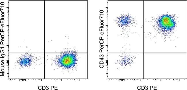

Invitrogen™ CD43 Monoclonal Antibody (4-29-5-10-21), PerCP-eFluor™ 710, eBioscience™, Invitrogen™


Description
CD43 (leukosialin, sialophorin) is a transmembrane mucin-like protein with high negative charge, expressed on the surface of most hematopoietic cells. CD43 contributes to a repulsive barrier that interferes with cellular adhesion, however, in certain cases also promotes leukocyte aggregation. By interaction with actin-binding proteins ezrin and moesin CD43 plays a regulatory role in remodeling T-cell morphology and regulates cell-cell interactions during lymphocyte traffic. CD43 signaling both enhances LFA-1 adhesiveness and counteracts LFA-1 induction via other receptors. Expression of CD43 causes induction of functionally active tumour suppressor p53 protein, but in case of p53 and ARF defficiency CD43 promotes tumour proliferation and viability. It appears to be an important modulator of leukocyte functions.
Specifications
Specifications
| Antigen | CD43 |
| Applications | Flow Cytometry |
| Classification | Monoclonal |
| Clone | 4-29-5-10-21 |
| Concentration | 5 μL/Test |
| Conjugate | PerCP-eFluor 710 |
| Formulation | PBS with BSA and 0.09% sodium azide; pH 7.2 |
| Gene | SPN |
| Gene Accession No. | P16150 |
| Gene Alias | 3E8 antigen; A630014B01Rik; B-cell differentiation antigen LP-3; CD43; CD43 cytoplasmic tail; CD43-ct; Galactoglycoprotein; GALGP; gpL115; Leukocyte sialoglycoprotein; leukosialin; leukosianin; LOC101120462; LOW QUALITY PROTEIN: leukosialin; Lsn; Lsn1; Ly48; Ly-48; Lymphocyte antigen 48; sialophorin; sialophorin (gpL115, leukosialin, CD43); sialophorin gpL115; SPN; W3/13 antigen |
| Show More |
Frequently Asked Questions (FAQs)
Our options will depend on the samples you are analyzing.
If cell viability is not critical, you can store your stained samples at 4 degrees C or on ice overnight in the dark and analyze the following day.
For samples stained with eFluor organic fluorochromes, we recommend that cells be suspended in 100 uL of Flow Cytometry Staining Buffer (Cat. No. 00-4222) and 100 uL of eBioscience IC Fixation Buffer (Cat. No. 00-8222); samples can be incubated for up to 3 days at 4 degrees C in the dark. Alternatively, the 1-step Fix/Lyse Solution (Cat. No. 00-5333) can be used. This is a great option when working with whole blood but also works for other cell types.
The eFluor Organic Dyes (eFluor 450, APC-eFluor 780, PerCP-eFluor 710, eFluor 710) are conventional fluorochromes. In contrast, the eVolve line of products are Quantum dots.
For Research Use Only.
By clicking Submit, you acknowledge that you may be contacted by Fisher Scientific in regards to the feedback you have provided in this form. We will not share your information for any other purposes. All contact information provided shall also be maintained in accordance with our Privacy Policy.