Promotional price valid on web orders only. Your contract pricing may differ. Interested in signing up for a dedicated account number?
Learn More
Learn More
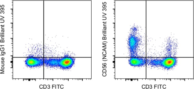
Invitrogen™ CD56 (NCAM) Monoclonal Antibody (TULY56), Brilliant Ultra Violet™ 395, eBioscience™
Mouse Monoclonal Antibody
Supplier: Invitrogen™ 363056641


Description
Description: This TULY56 monoclonal antibody reacts with human CD56, also known as Neural Cell Adhesion Molecule (NCAM). CD56 is a highly glycosylated transmembrane molecule expressed by neurons and plays a role in the homotypic adhesion of neural cells. In the hematopoietic system, CD56 is expressed on NK cells and a subset of T cells referred to as NKT cells. Staining with TULY56 does not block binding of CMSSB, suggesting that the two antibodies recognize different epitopes. Additionally, TULY56 performs better after fixation and permeabilization than CMSSB. The TULY56 monoclonal antibody crossreacts with Rhesus macaque. Applications Reported: This TULY56 antibody has been reported for use in flow cytometric analysis. Applications Tested: This TULY56 antibody has been pre-diluted and tested by flow cytometric analysis of normal human peripheral blood cells. This may be used at 5 μL (0.25 μg) per test. A test is defined as the amount (μg) of antibody that will stain a cell sample in a final volume of 100 μL. Cell number should be determined empirically but can range from 10^5 to 10^8 cells/test. Brilliant Ultra Violet™ 395 (BUV395) is a dye that emits at 395 nm and is intended for use on cytometers equipped with an ultraviolet (355 nm) laser. Please make sure that your instrument is capable of detecting this fluorochrome. When using two or more Super Bright, Brilliant Violet™, Brilliant Ultra Violet™, or other polymer dye-conjugated antibodi...
CD56, also known as neural cell adhesion molecule (NCAM), is a highly glycosylated transmembrane glycoprotein of the immunoglobulin family. It plays a crucial role in cell adhesion, migration, axonal growth, pathfinding, and synaptic plasticity. CD56 is ubiquitously expressed in the nervous system in isoforms ranging from 120-180 kDa and is involved in homotypic adhesion of neural cells. It mediates interactions by binding extracellular matrix components such as laminin and integrins, with polysialic modification reducing CD56-mediated adhesion. In the hematopoietic system, CD56 is expressed on natural killer (NK) cells and a subset of T cells known as NKT cells. It is also found on most neuroectodermal-derived cell lines, tissues, and neoplasms, including retinoblastoma, medulloblastoma, astrocytomas, and neuroblastoma. CD56 serves as a widely used neuroendocrine marker with high sensitivity for neuroendocrine tumors and ovarian granulosa cell tumors. Diseases associated with CD56 dysfunction include rabies and blastic plasmacytoid dendritic cell neoplasms, highlighting its importance in both neural and immune system functions.Specifications
| CD56 (NCAM) | |
| Monoclonal | |
| 5 μL/Test | |
| PBS with BSA and 0.09% sodium azide; pH 7.2 | |
| P13591 | |
| Ncam1 | |
| Affinity chromatography | |
| RUO | |
| 4684, 693789 | |
| 4°C, store in dark, DO NOT FREEZE! | |
| Liquid |
| Flow Cytometry | |
| TULY56 | |
| Brilliant Ultraviolet 395 | |
| Ncam1 | |
| adhesion molecule; antigen recognized by monoclonal antibody 5.1H11; Cd56; CD-56; CD56 120 kDa GPI-linked isoform; CD56 140 kDa isoform; CD56 140 kDa VASE isoform; E NCAM; E-NCAM; MSK39; N CAM1; NCAM; N-CAM; Ncam1; N-CAM-1; NCAM-1; NCAMC; NCAM-C; neural cell adhesion molecule; Neural cell adhesion molecule 1; neural cell adhesion molecule, NCAM; sCD56; sNCAM; soluble CD56; soluble NCAM | |
| Mouse | |
| 25 Tests | |
| Primary | |
| Human, Monkey, Rhesus Macaque | |
| Antibody | |
| IgG1 κ |
Product Content Correction
The Fisher Scientific Encompass Program offers items which are not part of our distribution portfolio. These products typically do not have pictures or detailed descriptions. However, we are committed to improving your shopping experience. Please use the form below to provide feedback related to the content on this product.
Product Title
Spot an opportunity for improvement?Share a Content Correction