Learn More
Invitrogen™ CD68 Monoclonal Antibody (815CU17), eFluor™ 570, eBioscience™


Description
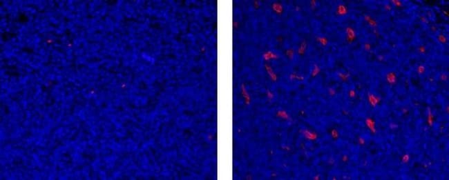
This 815CU17 monoclonal antibody reacts with human CD68, a 110 kDa, transmembrane glycoprotein that belongs to the sialomucin family and is closely related to the lysosomal-associated membrane proteins (LAMP) and scavenger receptor. CD68 is predominantly an intracellular protein, found mainly in the late endosomal compartment but can be detected in smaller amounts on the surface of mainly myeloid-derived cells: monocytes, macrophages, dendritic cells (and in some langerhans cells), neutrophils, basophils, mast cells, myeloid progenitor cells, and a subset of CD34+ hematopoietic bone marrow progenitor cells. The function has not been fully elucidated but based on homology and structure, CD68 may play a role in antigen processing or presentation and protection of the lysosomal membrane from hydrolytic enzymes. Reports have also shown expression in activated T cells, and about 40% of peripheral blood B lymphocytes and 50% of all B-ALL. This 815CU17 antibody does not cross-react with mouse or rat CD68. For flow cytometric analysis of human CD68, we recommend clone eBioY1/82A. Applications Reported: This 815CU17 antibody has been reported for use in microscopy and immunohistochemical staining of formalin-fixed paraffin embedded tissue sections. Applications Tested: This 815CU17 antibody has been tested by immunohistochemistry of formalin-fixed paraffin embedded human tissue using low or high pH antigen retreival and can be used at less than or equal to 10 ...
Specifications
Specifications
| Antigen | CD68 |
| Applications | Immunohistochemistry (Paraffin), Immunocytochemistry |
| Classification | Monoclonal |
| Clone | 815CU17 |
| Concentration | 0.2 mg/mL |
| Conjugate | eFluor 570 |
| Formulation | PBS with 0.09% sodium azide; pH 7.2 |
| Gene | Cd68 |
| Gene Accession No. | P34810 |
| Gene Alias | CD68; CD68 antigen; CD68 molecule; GP110; Lamp4; macrophage antigen CD68; macrosialin; Scard1; scavenger receptor class D, member 1; similar to CD68 antigen |
| Show More |
Frequently Asked Questions (FAQs)
Our options will depend on the samples you are analyzing.
If cell viability is not critical, you can store your stained samples at 4 degrees C or on ice overnight in the dark and analyze the following day.
For samples stained with eFluor organic fluorochromes, we recommend that cells be suspended in 100 uL of Flow Cytometry Staining Buffer (Cat. No. 00-4222) and 100 uL of eBioscience IC Fixation Buffer (Cat. No. 00-8222); samples can be incubated for up to 3 days at 4 degrees C in the dark. Alternatively, the 1-step Fix/Lyse Solution (Cat. No. 00-5333) can be used. This is a great option when working with whole blood but also works for other cell types.
Yes, the eFluor Organic fluorochromes can be used for intracellular staining. The eFluor organic fluorochromes maintain bright signal and require minimal changes in compensation when fixed with eBioscience IC Fixation Buffer (Cat. No. 00-8222-49) and Permeabilization Buffer (Cat. No. 00-8333-56) or 1-step Fix/Lyse Solution (Cat. No. 00-5333-54, 00-5333-57) (as compared to live cells).
The eFluor Organic Dyes (eFluor 450, APC-eFluor 780, PerCP-eFluor 710, eFluor 710) are conventional fluorochromes. In contrast, the eVolve line of products are Quantum dots.
As with other fluorochromes, we recommend minimal exposure to light to maintain optimal signal.
By clicking Submit, you acknowledge that you may be contacted by Fisher Scientific in regards to the feedback you have provided in this form. We will not share your information for any other purposes. All contact information provided shall also be maintained in accordance with our Privacy Policy.