Learn More
CD69 Mouse anti-Human, RB545, Clone: FN50 (also known as FN 50), BD Horizon™
Shop All BD Biosciences ProductsDescription
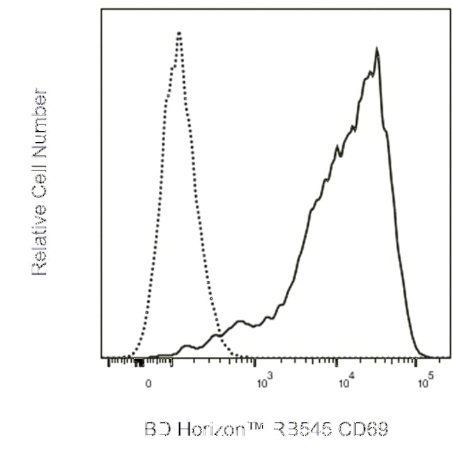
The FN50 monoclonal antibody specifically binds to human CD69. CD69 is also known as activation-induced molecule (AIM), early activation antigen (EA-1), very early activation antigen (VEA), C-type lectin domain family 2 member C (CLEC2C), MLR-3, GP32/28 and Leu-23. CD69 is a transmembrane type II homodimer receptor. CD69 is comprised of disulfide-linked, differentially glycosylated core protein subunits that are approximately 28 and 34 kDa in size. Each subunit contains a C-type lectin domain. CD69 is expressed on activated T, B, and natural killer (NK) lymphocytes, thymocytes, neutrophils, eosinophils and platelets. In normal peripheral blood, a small and variable percentage of lymphocytes typically express detectable membrane CD69 antigen. Upon activation, CD69 antigen expression increases on lymphocytes. Peak CD69 expression generally occurs within 18 hours of activation, preceding the appearance of HLA-DR, IL-2Rα (CD25) and transferrin receptor (CD71). CD69 is highly expressed on the bright CD3+ subset of thymocytes. FN50 monoclonal antibody labels NK cells and most lymphocytes of the follicular mantle and perifollicular/interfollicular zone as well as germinal center T cells of lymph nodes and tonsils. Studies indicate that CD69 serves as a signaling receptor in the activation of a variety of cell types.
Specifications
Specifications
| Antigen | CD69 |
| Applications | Flow Cytometry |
| Classification | Monoclonal |
| Clone | FN50 (also known as FN 50) |
| Conjugate | RB545 |
| Formulation | Aqueous buffered solution containing ≤0.09% sodium azide. |
| Gene Alias | AIM; CLEC2C; EA1; GP32/28; Leu23; MLR-3; VEA; BL-AC/P26 |
| Host Species | Mouse |
| Immunogen | Anti-μ stimulated human B lymphocytes |
| Purification Method | Affinity Purified |
| Show More |
By clicking Submit, you acknowledge that you may be contacted by Fisher Scientific in regards to the feedback you have provided in this form. We will not share your information for any other purposes. All contact information provided shall also be maintained in accordance with our Privacy Policy.