Learn More
Invitrogen™ CD7 Monoclonal Antibody (eBio124-1D1 (124-1D1)), Biotin, eBioscience™, Invitrogen™
Mouse Monoclonal Antibody
Supplier: Invitrogen™ 13007982

Description
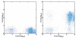
Description: The eBio124-1D1 monoclonal antibody reacts with human CD7, also known as gp40 and Leu9. CD7, a 40 kD receptor, is a member of the immunoglobulin gene superfamily. The N-terminal amino acid sequence (aa1-107) is highly homologous to Ig kappa light chain sequence; while the carboxyl-terminal region of the extracellular domain is proline-rich and has been postulated to form a stalk from which the Ig domain projects. CD7 is expressed on the majority of immature and mature T lymphocytes, and T cell leukemias. It is also found on natural killer cells, a small suppopulation of normal B cells and on maligant B cells. Cross-linking surface CD7 positively modulates T cell and NK cell activity, as measured by calcium flux, expression of adhesion molecules, cytokine secretion and proliferation. CD7 associates directly with phosphoinositol 3'-kinase. CD7 ligation induces production of D-3 phosphoinositides and tyrosine phosphorylation. A clonogenic subpopulation of human CD34(+) CD38(-) cord blood cells that express CD45RA and HLA-DR and high levels of the CD7 has been reported. These cells possess the capacity for lymphopoiesis. They can generate B-cells, natural killer cells, and dendritic cells but do not possess the capacity to develop into myeloid cells or erythroid cells. The CD7(+) phenotype distinguishes primitive human lymphoid progenitors from pluripotent stem cells.
CD7, also known as gp40 or Leu9, is a 40 kDa receptor and member of the immunoglobulin gene superfamily. It features an N-terminal region (amino acids 1-107) that is highly homologous to Ig kappa light chains, while its carboxyl-terminal region is proline-rich, forming a stalk from which the Ig domain projects. CD7 is prominently expressed on the majority of immature and mature T lymphocytes, as well as T cell leukemias. It is also found on natural killer cells, a small subpopulation of normal B cells, and malignant B cells. CD7 plays a crucial role in modulating immune cell activity. Cross-linking of surface CD7 enhances T cell and NK cell functions, as evidenced by increased calcium flux, expression of adhesion molecules, cytokine secretion, and proliferation. CD7 directly associates with phosphoinositol 3-kinase, and its ligation induces the production of D-3 phosphoinositides and tyrosine phosphorylation. The expression of CD7 is an important marker in leukemia diagnostics, highlighting its significance in both normal immune function and disease states.
Specifications
| CD7 | |
| Monoclonal | |
| 0.5 mg/mL | |
| PBS with 0.09% sodium azide; pH 7.2 | |
| P09564 | |
| CD7 | |
| Affinity chromatography | |
| RUO | |
| 924 | |
| 4°C, store in dark, DO NOT FREEZE! | |
| Liquid |
| Flow Cytometry | |
| eBio124-1D1 (124-1D1) | |
| Biotin | |
| CD7 | |
| Cd7; CD7 antigen; CD7 antigen (p41); Cd7 molecule; GP40; LEU-9; p41 protein; T-cell antigen CD7; T-cell leukemia antigen; T-cell surface antigen Leu-9; Tp40; TP41 | |
| Mouse | |
| 100 μg | |
| Primary | |
| Human | |
| Antibody | |
| IgG1 κ |
Safety and Handling
The Fisher Scientific Encompass Program offers items which are not part of our distribution portfolio. These products typically do not have pictures or detailed descriptions. However, we are committed to improving your shopping experience. Please use the form below to provide feedback related to the content on this product.
For Research Use Only.
