Learn More
Invitrogen™ CD83 Monoclonal Antibody (Michel-17 (Michel17)), eFluor™ 660, eBioscience™
Rat Monoclonal Antibody
Supplier: Invitrogen™ 50083182

Description
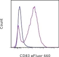
Description: The Michel-17 monoclonal antibody reacts with mouse CD83, a 45kDa cell surface glycoprotein and a member of the Ig superfamily. The mouse CD83 antigen is expressed predominantly on mature DC and activated lymphocytes. Cross-linking of CD83 with Michel-17 on DC or activated T cells does not induce any activation signal. CD83 plays an important role in T cell development through interaction with its ligand. CD83-Ig protein has revealed the presence of a CD83 ligand expressed mainly by B220+ cells in mouse spleen. Applications Reported: This Michel-17 (Michel17) antibody has been reported for use in flow cytometric analysis. Applications Tested: This Michel-17 (Michel17) antibody has been tested by flow cytometric analysis of stimulated mouse splenocytes. This can be used at less than or equal to 0.25 μg per test. A test is defined as the amount (μg) of antibody that will stain a cell sample in a final volume of 100 μL. Cell number should be determined empirically but can range from 10^5 to 10^8 cells/test. It is recommended that the antibody be carefully titrated for optimal performance in the assay of interest. eFluor® 660 is a replacement for Alexa Fluor® 647. eFluor® 660 emits at 659 nm and is excited with the red laser (633 nm). Please make sure that your instrument is capable of detecting this fluorochome. Excitation: 633-647 nm; Emission: 668 nm; Laser: Red Laser. Filtration: 0.2 μm post-manufacturing filtered.
CD83 cell surface antigen is a 40-45kD glycoprotein expressed by peripheral blood dendritic cells. Peripheral lymphocytes can be induced to express very low levels of CD83 after culture in agents such as Con A or PHA. In immunohistology, CD83 is shown to be expressed strongly by interfollicular interdigitating reticulum cells and more weakly by cells within germinal centres. CD83 is also expressed by Langerhan's cells in the skin. The CD83 antigen is a 186-amino-acid single-chain glycoprotein and this molecule is a member of the immunoglobulin superfamily that is composed of an extracellular V-type Ig-like single domain, a transmembrane region, and a short, 40-amino-acid cytoplasmic tail. CD83 antigen undergoes extensive post-translational glycosylation, since the determined Mr is twice the predicted size of the core protein. However, CD83+ cells have a unique cell surface immuno-phenotype that does not correlate with that of T cells, B cells, NK cells, or cells of the myelomonocytic lineage. CD83+ cells coexpress the highest levels of MHC class II molecules, when compared with other leucocyte lineages. They also co-express T cell markers (CD2, CD5), B cell markers (CD40, CD78), myeloid cell markers (CD13, CD33, CD36), cytokine receptors as well as other cell surface molecules. Diseases associated with CD83 dysfunction include plague and Rift Valley Fever.
Specifications
| CD83 | |
| Monoclonal | |
| 0.2 mg/mL | |
| PBS with 0.09% sodium azide; pH 7.2 | |
| O88324 | |
| CD83 | |
| Affinity chromatography | |
| RUO | |
| 12522 | |
| 4°C, store in dark, DO NOT FREEZE! | |
| Liquid |
| Flow Cytometry | |
| Michel-17 (Michel17) | |
| eFluor 660 | |
| CD83 | |
| B-cell activation protein; BL11; CD antigen CD83; CD83; CD83 antigen; CD83 antigen (activated B lymphocytes, immunoglobulin superfamily); CD83 molecule; cell surface protein HB15; cell-surface glycoprotein; HB15; hCD83; mCD83 | |
| Rat | |
| 100 μg | |
| Primary | |
| Mouse | |
| Antibody | |
| IgG1 |
The Fisher Scientific Encompass Program offers items which are not part of our distribution portfolio. These products typically do not have pictures or detailed descriptions. However, we are committed to improving your shopping experience. Please use the form below to provide feedback related to the content on this product.

