Promotional price valid on web orders only. Your contract pricing may differ. Interested in signing up for a dedicated account number?
Learn More
Learn More
Invitrogen™ Claudin 2 Recombinant Superclonal™ Antibody (3HCLC)
Rabbit Recombinant Superclonal Antibody
Supplier: Invitrogen™ 710221
Description
This antibody is predicted to react with equine, mouse and rat based on sequence homology. Recombinant rabbit Superclonal™ antibodies are unique offerings from Thermo Fisher Scientific. They are comprised of a selection of multiple different recombinant monoclonal antibodies, providing the best of both worlds - the sensitivity of polyclonal antibodies with the specificity of monoclonal antibodies - all delivered with the consistency only found in a recombinant antibody. While functionally the same as a polyclonal antibody - recognizing multiple epitope sites on the target and producing higher detection sensitivity for low abundance targets - a recombinant rabbit Superclonal™ antibody has a known mixture of light and heavy chains. The exact population can be produced in every lot, circumventing the biological variability typically associated with polyclonal antibody production. Note: Formerly called Recombinant polyclonal antibody, this product is now rebranded as Recombinant Superclonal™ antibody. The physical product and the performance remain unchanged.
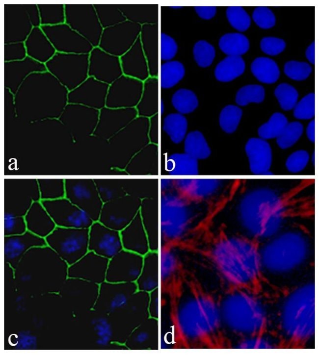
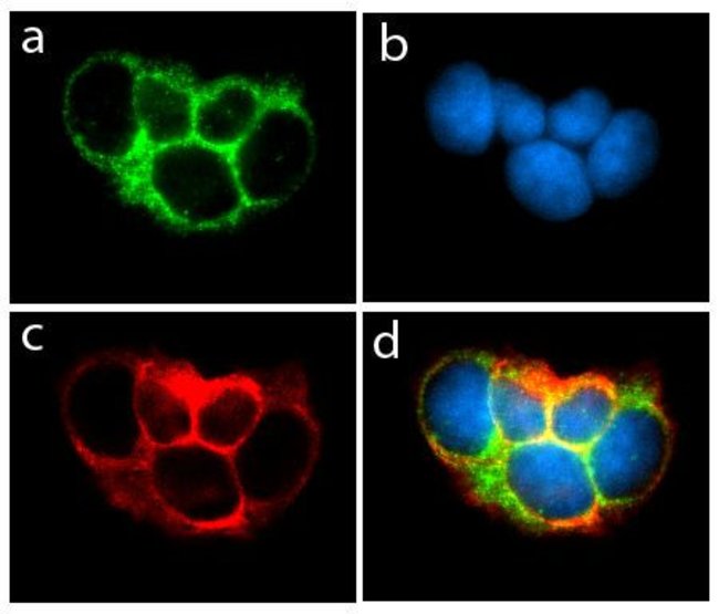
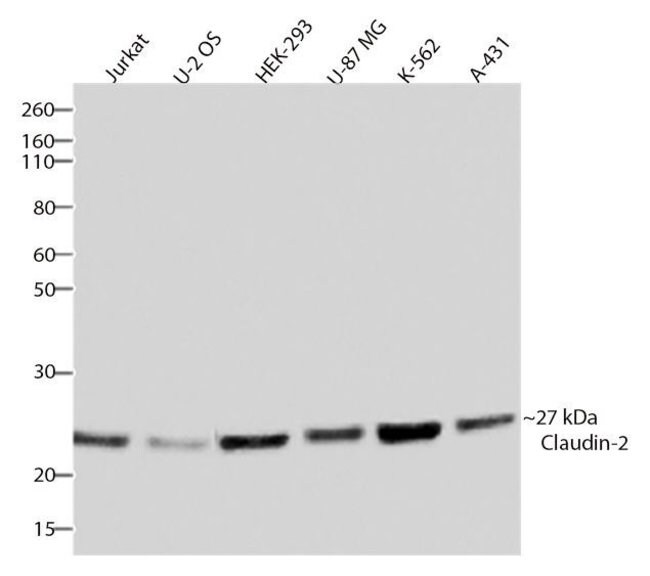
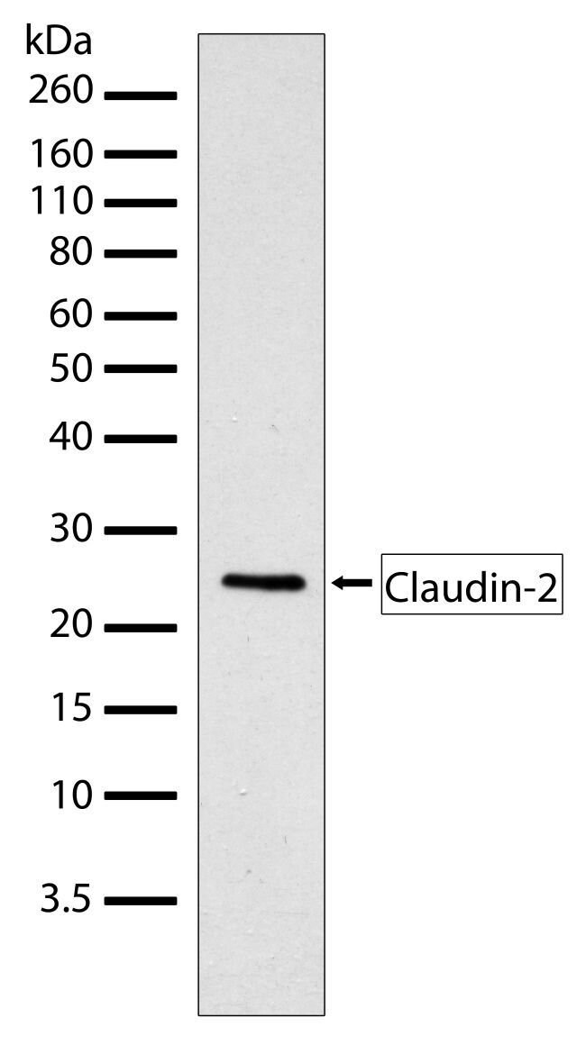
Tight junctions are specialized regions of cell-cell contact that are particularly abundant in luminal epithelial cell sheets. In freeze-fracture electron micrographs, tight junctions are visualized as belt-like bands of anastomosing sealing strands (TJ strands) that completely encircle the lateral surfaces of each cell. TJ strands on adjacent cells are presumed to interact with each other to form a sort of molecular gasket that prevents ions, water and other molecules from leaking between cells and thus, from one side of the sheet to the other. In addition to this so-called barrier function, the fence function of tight junctions plays an important role in maintaining epithelial cell-polarity by blocking the diffusion of membrane proteins between apical (luminal) and basolateral cell surfaces. Confinement of, for example, the glucose symport to apical surfaces allows glucose to be transported vectorially from the lumen, through the cell, and into the bloodstream. Several peripheral membrane proteins are associated with tight junctions including ZO-1, ZO-2, ZO-3 (members of membrane-associated guanylate-kinase family), cingulin, the 7H6 antigen, Rab-3b, symplekin. While their precise functions are not known, roles for these proteins have been suggested in tight junction assembly and maintenance; signal transduction; and the regulation of tight junction permeability. Furthermore, a growing body of evidence suggests that actin filaments play a major role in regulating tight junction permeability. Until recently, the only transmembrane protein known to be associated with tight junctions was occludin, an ~65 kDa protein with four transmembrane domains. Despite widespread expectation, a critical structural role for occludin in TJ strands was ruled out by the observation of apparently normal tight junctions formed between cells disrupted at both occludin alleles. Fortunately, a closer examination of isolated tight junctions uncovered two related ~22 kDa, four-transmembrane domain proteins, claudin-1 and claudin-2, with no similarity to occludin. In contrast to occludin, which induces only a small number of short strands at cell-cell contact sites when introduced into fibroblasts lacking tight junctions, claudin-1 and -2 induce networks of strands characteristic of true tight junctions. Though inconclusive, these findings suggest that claudin-1 and -2 are major structural components of TJ strands and that occludin plays some other accessory role. Excitement in the tight junction field continues to rise following the recent discovery of claudins -3, -4, -5, -6, -7, and -8 and experiments suggesting that tight junctions in different tissues are comprised of different sets of claudin family proteins. Claudin-1 and claudin-2 connect to the actin cytoskeleton through ZO-1; Claudin-2 functions as a paracellular channel with cation (Na+) selectivity at tight junctions. The expression of claudin-2 is restricted to the liver and kidney, with small amounts also found in the brain.Specifications
| Claudin 2 | |
| Recombinant Superclonal | |
| 0.5 mg/mL | |
| PBS with 0.09% sodium azide | |
| P57739 | |
| CLDN2 | |
| Peptide corresponding to amino acids 188-230 of human claudin-2. | |
| 100 μg | |
| Primary | |
| Human | |
| Antibody | |
| IgG |
| Western Blot, Immunocytochemistry | |
| 3HCLC | |
| Unconjugated | |
| CLDN2 | |
| AL022813; claudin 2; Claudin2; claudin-2; CLD2; Cldn2; integral membrane protein claudin-2; PSEC0059; RGD1560247; SP82; UNQ705/PRO1356 | |
| Rabbit | |
| Protein A | |
| RUO | |
| 9075 | |
| Store at 4°C short term. For long term storage, store at -20°C, avoiding freeze/thaw cycles. | |
| Liquid |
Safety and Handling
WARNING: Cancer - www.P65Warnings.ca.gov
Product Content Correction
Your input is important to us. Please complete this form to provide feedback related to the content on this product.
Product Title
Spot an opportunity for improvement?Share a Content Correction