Learn More
Invitrogen™ CXCL9 (MIG) Monoclonal Antibody (MIG-2F5.5), eFluor™ 660, eBioscience™


Description
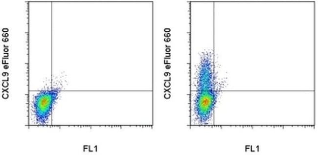
Description: The monoclonal antibody MIG-2F5.5 reacts with mouse monokine induced by IFN gamma (mig, mig-1, CXCL9). This 14.4 kDa inflammatory chemokine is specifically induced by IFN gamma, but not other activators such as LPS or IFN alpha. Secretion of mig, mainly by macrophages results in the chemotaxis of a variety of activated T cells via the CXCR3 chemokine receptor. Mig is involved many areas of research including autoimmune diseases, cancer, and inflammation. Applications Reported: This MIG-2F5.5 antibody has been reported for use in intracellular staining followed by flow cytometric analysis. Applications Tested: This MIG-2F5.5 antibody has been tested by intracellular staining and flow cytometric analysis of stimulated RAW 264.7 cells. This can be used at less than or equal to 0.25 μg per test. A test is defined as the amount (μg) of antibody that will stain a cell sample in a final volume of 100 μL. Cell number should be determined empirically but can range from 10^5 to 10^8 cells/test. It is recommended that the antibody be carefully titrated for optimal performance in the assay of interest. eFluor® 660 is a replacement for Alexa Fluor® 647. eFluor® 660 emits at 659 nm and is excited with the red laser (633 nm). Please make sure that your instrument is capable of detecting this fluorochome. Excitation: 633-647 nm; Emission: 668 nm; Laser: Red Laser. Filtration: 0.2 μm post-manufacturing filtered.

Specifications
Specifications
| Antigen | CXCL9 (MIG) |
| Applications | Flow Cytometry |
| Classification | Monoclonal |
| Clone | MIG-2F5.5 |
| Concentration | 0.2 mg/mL |
| Conjugate | eFluor 660 |
| Formulation | PBS with 0.09% sodium azide; pH 7.2 |
| Gene | CXCL9 |
| Gene Accession No. | P18340 |
| Gene Alias | BB139920; C Cmotif chemokine; C X C motif chemokine; CC motif chemokine; CCmotif chemokine; chemokine (C-X-C motif) ligand 9; CMK; crg-10; CXC; CXC motif chemokine; C-X-C motif chemokine 9; C-X-C motif chemokine ligand 9; CXCL; CXCL9; gamma interferon-induced monokine; gamma-interferon-induced monokine; HuMIG; M119; Mig; monokine induced by gamma interferon; monokine induced by interferon gamma; monokine induced by interferon-gamma; MuMIG; protein m119; SCYB9; small inducible cytokine B subfamily (Cys-X-Cys), member 9; small inducible cytokine B9; small-inducible cytokine B9 |
| Show More |
Frequently Asked Questions (FAQs)
Our options will depend on the samples you are analyzing.
If cell viability is not critical, you can store your stained samples at 4 degrees C or on ice overnight in the dark and analyze the following day.
For samples stained with eFluor organic fluorochromes, we recommend that cells be suspended in 100 uL of Flow Cytometry Staining Buffer (Cat. No. 00-4222) and 100 uL of eBioscience IC Fixation Buffer (Cat. No. 00-8222); samples can be incubated for up to 3 days at 4 degrees C in the dark. Alternatively, the 1-step Fix/Lyse Solution (Cat. No. 00-5333) can be used. This is a great option when working with whole blood but also works for other cell types.
Yes, in-house studies have demonstrated that the eFluor 660 fluorochrome is recognized by Anti-Cy5/Alexa Fluor 647 beads. Side by side studies with Alexa Fluor 647 versus eFluor 660 conjugated antibodies have demonstrated comparable results.
The eFluor Organic fluorochromes and eVolve QDots can be used with flow staining buffers containing PBS and protein.
The eFluor Organic Dyes (eFluor 450, APC-eFluor 780, PerCP-eFluor 710, eFluor 710) are conventional fluorochromes. In contrast, the eVolve line of products are Quantum dots.
As with other fluorochromes, we recommend minimal exposure to light to maintain optimal signal.
By clicking Submit, you acknowledge that you may be contacted by Fisher Scientific in regards to the feedback you have provided in this form. We will not share your information for any other purposes. All contact information provided shall also be maintained in accordance with our Privacy Policy.