Promotional price valid on web orders only. Your contract pricing may differ. Interested in signing up for a dedicated account number?
Learn More
Learn More

Cytokeratin, pan Antibody (AE-1/AE-3) - Azide and BSA Free, Novus Biologicals™

Mouse Monoclonal Antibody has been used in 4 publications
$658.00
Specifications
| Antigen | pan Cytokeratin |
|---|---|
| Clone | AE-1/AE-3 |
| Concentration | 1.0 mg/mL |
| Dilution | Western Blot 0.5-1ug/ml, Flow Cytometry 0.1 - 1 ug/million cells, Immunohistochemistry, Immunocytochemistry/Immunofluorescence 1-2ug/ml, Immunohistochemistry-Paraffin 0.25 - 0.5 ug/ml, Immunohistochemistry-Frozen 0.5-1ug/mlimmunohistochemistry-Paraffin 0.5-1ug/ml, Flow (Intracellular), CyTOF-ready, Dual RNAscope ISH-IHC |
| Applications | Western Blot, Flow Cytometry, Immunohistochemistry, Immunocytochemistry, Immunofluorescence, Immunohistochemistry (Paraffin), Immunohistochemistry (Frozen), CyTOF |
Description
Cytokeratin, pan Monoclonal specifically detects Cytokeratin, pan in Human, Mouse, Rat, Bovine, Canine, Chicken, Monkey, Rabbit, Reptile, Zebrafish samples. It is validated for Western Blot, Flow Cytometry, Immunohistochemistry, Immunocytochemistry/Immunofluorescence, Immunohistochemistry-Paraffin, Immunohistochemistry-Frozen, Flow (Intracellular), CyTOF-ready, Single Cell Western, Dual RNAscope ISH-IHC.Specifications
| pan Cytokeratin | |
| 1.0 mg/mL | |
| Western Blot, Flow Cytometry, Immunohistochemistry, Immunocytochemistry, Immunofluorescence, Immunohistochemistry (Paraffin), Immunohistochemistry (Frozen), CyTOF | |
| Unconjugated | |
| Mouse | |
| Apoptosis, Cancer, Cell Biology, Cellular Markers, Signal Transduction | |
| 3848 | |
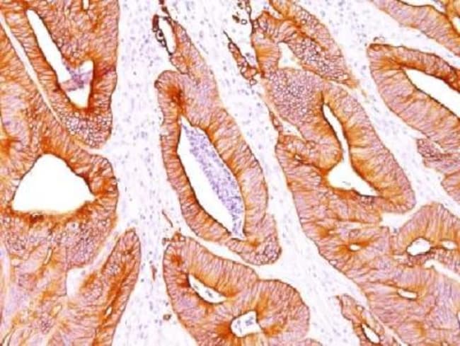
| This Cytokeratin, pan Antibody (AE-1/AE-3) was developed against total keratin isolated from human epidermal callus was used as immunogen to generate the pan cytokeratin antibodies AE1 + AE3 (Woodcock-Mitchell, 1982). | |
| Protein A or G purified | |
| Twenty human keratins are resolved with two-dimensional gel electrophoresis into acidic (pI 6.0) subfamilies. This antibody cocktail recognizes acidic (Type I or LMW) and basic (Type II or HMW) cytokeratins, which 67kDa (CK1); 64kDa (CK3); 59kDa (CK4); 58kDa (CK5); 56kDa (CK6); 52kDa (CK8); 56.5kDa (CK10); 50kDa (CK14); 50kDa (CK15); 48kDa (CK16); 40kDa (CK19). Many studies have shown the usefulness of keratins as markers in cancer research and tumor diagnosis. AE-1/AE-3 is a broad spectrum anti pan-cytokeratin antibody cocktail, which differentiates epithelial tumors from non-epithelial tumors e.g. squamous vs. adenocarcinoma of the lung, liver carcinoma, breast cancer, and esophageal cancer. It has been used to characterize the source of various neoplasms and to study the distribution of cytokeratin containing cells in epithelia during normal development and during the development of epithelial neoplasms. This antibody stains cytokeratins present in normal and abnormal human tissues and has shown high sensitivity in the recognition of epithelial cells and carcinomas. |
| AE-1/AE-3 | |
| Western Blot 0.5-1ug/ml, Flow Cytometry 0.1 - 1 ug/million cells, Immunohistochemistry, Immunocytochemistry/Immunofluorescence 1-2ug/ml, Immunohistochemistry-Paraffin 0.25 - 0.5 ug/ml, Immunohistochemistry-Frozen 0.5-1ug/mlimmunohistochemistry-Paraffin 0.5-1ug/ml, Flow (Intracellular), CyTOF-ready, Dual RNAscope ISH-IHC | |
| Monoclonal | |
| Purified | |
| RUO | |
| Human, Mouse, Rat, Bovine, Canine, Chicken, Primate, Rabbit, Reptile, Zebrafish | |
| KRT1 | |
| Primary | |
| Store at 4C short term. Aliquot and store at -20C long term. Avoid freeze-thaw cycles. |
For Research Use Only
Spot an opportunity for improvement?Share a Content Correction
Product Content Correction
Your input is important to us. Please complete this form to provide feedback related to the content on this product.
Product Title