Learn More

Invitrogen™ Desmin Monoclonal Antibody (DE-U-10), eFluor™ 660, eBioscience™


Description
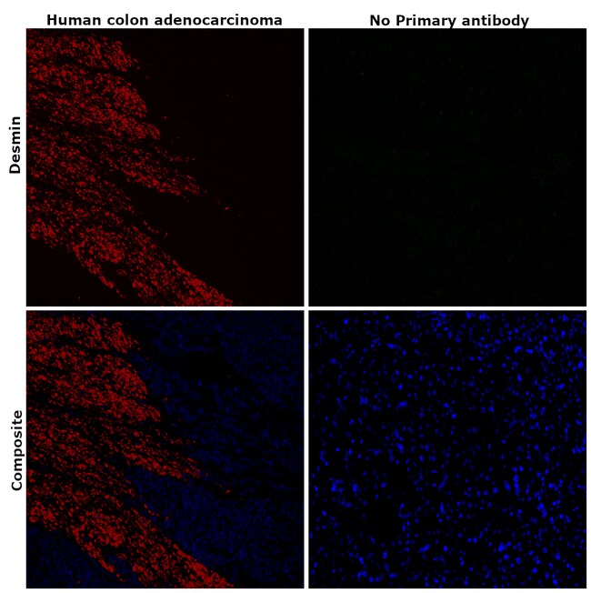
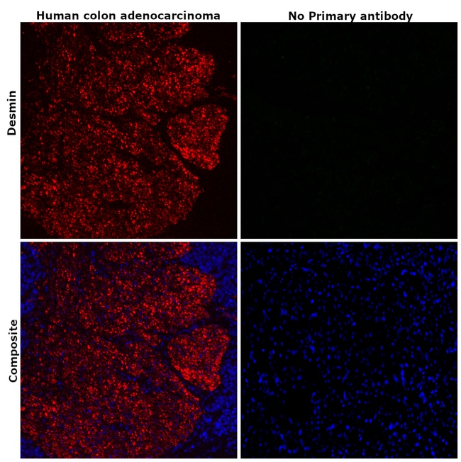
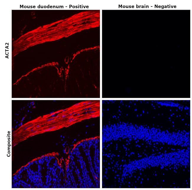
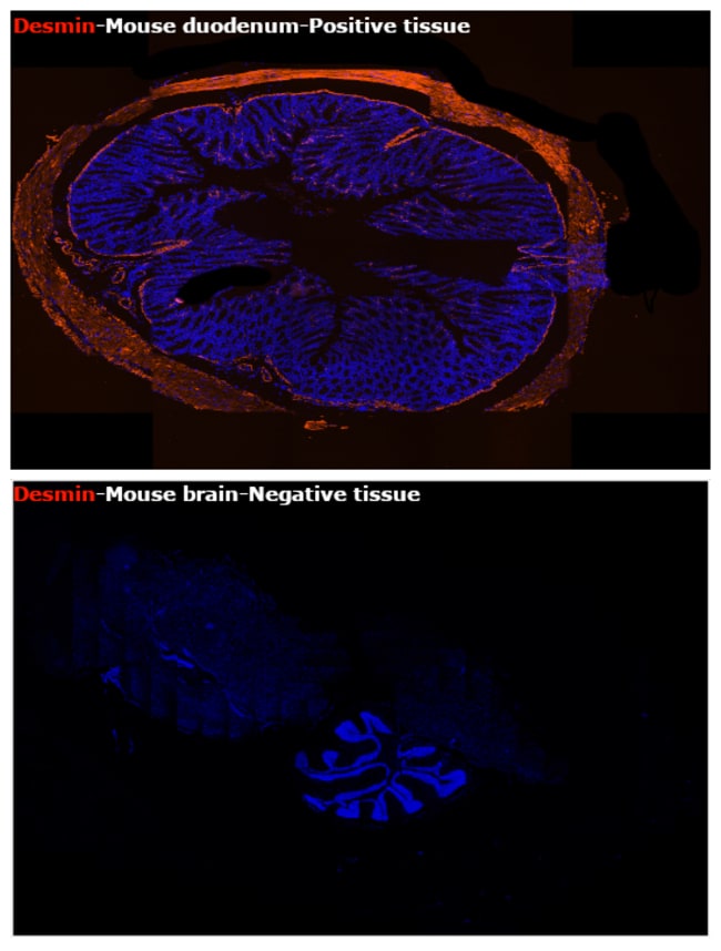
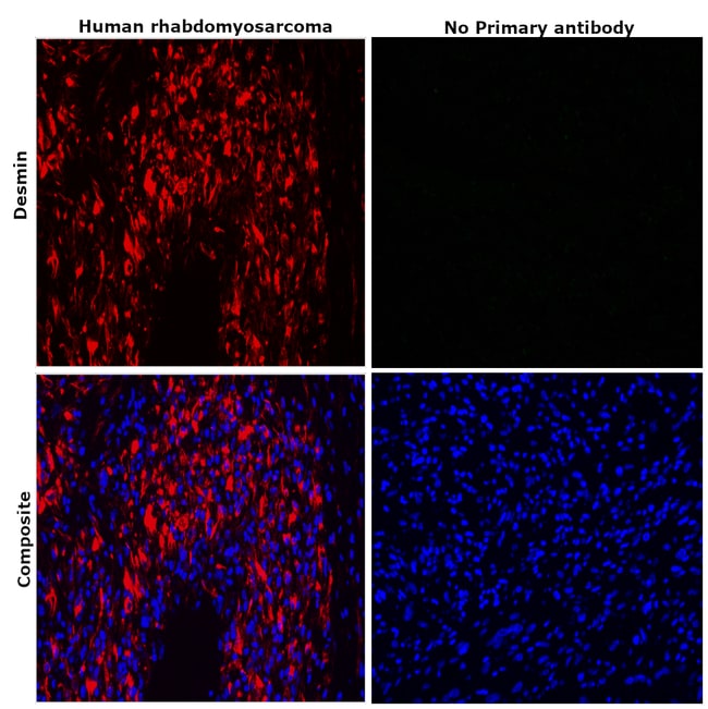
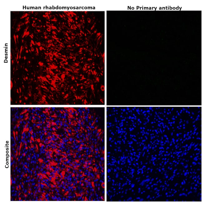
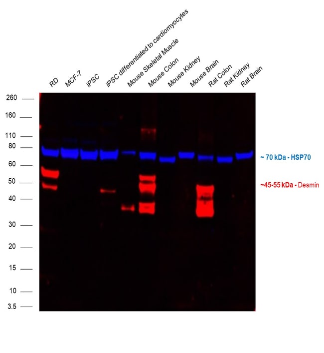
Description: The monoclonal antibody DE-U-10 recognizes desmin. Desmin is the protein subunit of class-III intermediate filaments. Desmin is found predominantly in skeletal, cardiac, and smooth muscle. Desmin plays a role in muscle cell development and differentiation, muscle cell architecture and structure, and mitochondrial function. Desmin forms scaffolds around the Z-disk of sarcomeres in muscle cells. Desmin knockout mice exhibit defects in skeletal, smooth, and cardiac muscle and have impaired mitochondrial function. Mutations in Desmin result in conditions such as desmin-related myopathy (DRM), cardiomyopathy dilated type 1I (CMD1I), and neurogenic scapuloperoneal syndrome Kaeser type (Kaeser syndrome). The DE-U-10 antibody recognizes human, mouse, rat, feline, bovine, goat, hamster, sheep, porcine, rabbit, and chicken desmin. The DE-U-10 antibody will also label tumors derived from muscle tissue, such as leiomyomas and rhabdomyosarcomas. Applications Reported: This DE-U-10 antibody has been reported for use in immunohistochemical staining of formalin-fixed paraffin embedded tissue sections, microscopy, and immunocytochemistry. Applications Tested: This DE-U-10 antibody has been tested by immunohistochemistry of formalin-fixed paraffin embedded human tissue using high pH antigen retrieval and can be used at less than or equal to 10 μg/mL. This DE-U-10 antibody has been tested by immunocytochemistry of fixed and permeabilized cells and can be used at less than...
Specifications
Specifications
| Antigen | Desmin |
| Applications | Immunohistochemistry (Paraffin), Western Blot, Immunocytochemistry |
| Classification | Monoclonal |
| Clone | DE-U-10 |
| Concentration | 0.2 mg/mL |
| Conjugate | eFluor 660 |
| Formulation | PBS with 0.09% sodium azide; pH 7.2 |
| Gene | DES |
| Gene Accession No. | O62654, P02540, P02542, P17661, P31001, P48675 |
| Gene Alias | CMD1I; CSM1; CSM2; DES; Desmin; desmin-like protein; FLJ12025; FLJ39719; FLJ41013; FLJ41793; I79_018932; intermediate filament protein; LGMD2R; MFM1; muscle-specific intermediate filament desmin; muscle-specific intermediate filament protein; mutant desmin p.K241E; SCPNK; similar to desmin |
| Show More |
Frequently Asked Questions (FAQs)
Our options will depend on the samples you are analyzing.
If cell viability is not critical, you can store your stained samples at 4 degrees C or on ice overnight in the dark and analyze the following day.
For samples stained with eFluor organic fluorochromes, we recommend that cells be suspended in 100 uL of Flow Cytometry Staining Buffer (Cat. No. 00-4222) and 100 uL of eBioscience IC Fixation Buffer (Cat. No. 00-8222); samples can be incubated for up to 3 days at 4 degrees C in the dark. Alternatively, the 1-step Fix/Lyse Solution (Cat. No. 00-5333) can be used. This is a great option when working with whole blood but also works for other cell types.
Yes, the eFluor Organic fluorochromes can be used for intracellular staining. The eFluor organic fluorochromes maintain bright signal and require minimal changes in compensation when fixed with eBioscience IC Fixation Buffer (Cat. No. 00-8222-49) and Permeabilization Buffer (Cat. No. 00-8333-56) or 1-step Fix/Lyse Solution (Cat. No. 00-5333-54, 00-5333-57) (as compared to live cells).
Yes, in-house studies have demonstrated that the eFluor 660 fluorochrome is recognized by Anti-Cy5/Alexa Fluor 647 beads. Side by side studies with Alexa Fluor 647 versus eFluor 660 conjugated antibodies have demonstrated comparable results.
The eFluor Organic Dyes (eFluor 450, APC-eFluor 780, PerCP-eFluor 710, eFluor 710) are conventional fluorochromes. In contrast, the eVolve line of products are Quantum dots.
As with other fluorochromes, we recommend minimal exposure to light to maintain optimal signal.
By clicking Submit, you acknowledge that you may be contacted by Fisher Scientific in regards to the feedback you have provided in this form. We will not share your information for any other purposes. All contact information provided shall also be maintained in accordance with our Privacy Policy.