Promotional price valid on web orders only. Your contract pricing may differ. Interested in signing up for a dedicated account number?
Learn More
Learn More
MilliporeSigma™ Dystrophin, Mouse, Alexa Fluor 488, Clone: 2C6 (MANDYS106) ,
Mouse Monoclonal Antibody
Supplier: MilliporeSigma™ MABT827AF488
Description
Dystrophin (UniProt P11532) is encoded by the DMD (also known as BMD, CMD3B, DXS142, DXS164, DXS206, DXS230, DXS239, DXS268, DXS269, DXS270, DXS272, MRX85) gene (Gene ID 1756) in human. Dystrophin is localized to the inner part of the muscle fiber cell membrane (sarcolemma), where it forms the dystrophin-associated glycoprotein complex (DGC) that links the extracellular matrix to the actin cytoskeleton. Dystrophin plays an important role in stabilizing the muscle fibre against the mechanical forces of muscle contraction by providing a shock-absorbing connection between the cytoskeleton and the extracellular matrix. Duchenne muscular dystrophy (DMD) is caused by gene mutations that disrupt the open reading frame (ORF) and prevent the full translation of dystrophin. ORF restoration by exon skipping using antisense oligonucleotides targeted to splicing elements are designed to transform the DMD phenotype to that of the milder disorder, Becker muscular dystrophy (BMD), typically caused by in-frame dystrophin deletions that allow the production of an internally deleted but partially functional dystrophin.
Specifications
| Dystrophin | |
| Monoclonal | |
| Please refer to lot specific datasheet. | |
| Purified mouse IgG2a Alexa Fluor™ 488 conjugate in buffer containing PBS, 1.5% BSA with 0.05% Sodium Azide. | |
| P11532 | |
| TrpE-tagged recombinant protein corresponding to the Exon 43-coded pectrin-like repeat 16 region of human Dystrophin. | |
| 100 μL | |
| Cell Structure | |
| NP_000100 | |
| Human | |
| IgG2a κ |
| Immunofluorescence | |
| 2C6 (MANDYS106) | |
| Alexa Fluor 488 | |
| DMD, BMD, CMD3B, DXS142, DXS164, DXS206, DXS230, DXS239, DXS268, DXS269, DXS270, DXS272, MRX85 | |
| Mouse | |
| Protein G Purified | |
| RUO | |
| Primary | |
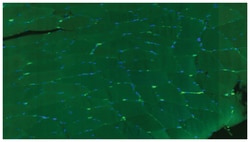
| Detects dystrophin spliced isoforms 1-4, but not isoforms 5-10, or utrophin. Positive muscle membrane staining of tissue samples from healthy donors, reduced staining of Becker muscular dystrophy (BMD) biopsies, and no staining is seen with Duchenne muscular dystrophy (DMD) biopsy samples (Anthony, K., et al. (2011). Brain. 134(Pt 12):3547-3559). | |
| Stable for 1 year at 2°-8°C from date of receipt. |
Product Content Correction
Your input is important to us. Please complete this form to provide feedback related to the content on this product.
Product Title
For Research Use Only. Not for use in diagnostic procedures.
Spot an opportunity for improvement?Share a Content Correction
