Promotional price valid on web orders only. Your contract pricing may differ. Interested in signing up for a dedicated account number?
Learn More
Learn More
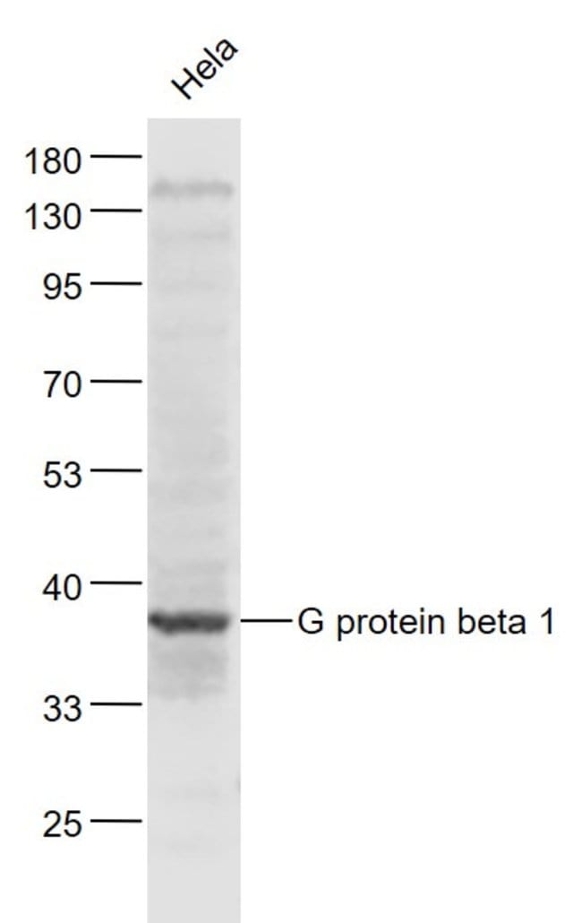
G protein beta 1/GNB1 Rabbit anti-Human, Polyclonal, Bioss


Description
Vision involves the conversion of light into electrochemical signals that are processed by the retina and subsequently sent to and interpreted by the brain. The process of converting light into an electrochemical signal begins when the membrane-bound protein, rhodopsin, absorbs light within the retina. Photoexcitation of rhodopsin causes the cytoplasmic surface of the protein to become catalytically active. In the active state, rhodopsin activates transducin, a GTP binding protein. Once activated, transducin promotes the hydrolysis of cGMP by phosphodiesterase (PDE). The decrease of intracellular cGMP concentration causes the ion channels within the outer segment of the rod or cone to close, thus causing membrane hyperpolarization and, eventually, signal transmission. Rhodopsin activity is believed to be shut off by phosphorylation followed by binding of the soluble protein, arrestin. Transducin, once activated by rhodopsin, promotes the hydrolysis of cGMP by PDE. The subunit composition of transducin differs between different photoreceptor cells. Rod transducin consists of rod transducin alpha (Tr alpha), T beta, and T gamma. Cone transducin is composed of cone transducin alpha (Tc alpha), T beta and T gamma. Differential transducin subunit composition of transducin is believed to be responsible for the different light sensitivities between photoreceptive cells.
Specifications
Specifications
| Antigen | G protein beta 1/GNB1 |
| Applications | Immunofluorescence, Immunohistochemistry (Paraffin), Western Blot |
| Classification | Polyclonal |
| Concentration | 1 μg/mL |
| Conjugate | Unconjugated |
| Formulation | PBS with 1% BSA, 50% glycerol and 0.09% sodium azide; pH |
| Gene | GNB1 |
| Gene Accession No. | P62873 |
| Gene Alias | AA409223; beta subunit, signal-transducing proteins GS/GI; BOS_15860; C77571; G protein subunit beta 1; GNB1; Gnb-1; gnb1 protein; gnb1a; guanine nucleotide binding protein (G protein), beta 1; guanine nucleotide binding protein (G protein), beta polypeptide 1; guanine nucleotide binding protein (G protein), beta polypeptide 1a; guanine nucleotide binding protein, beta 1; Guanine nucleotide-binding protein beta 1; Guanine nucleotide-binding protein G(I)/G(S)/G(T) subunit beta-1; guanine nucleotide-binding protein, beta-1 subunit; MRD42; T beta; testicular tissue protein Li 72; Transducin beta chain 1; wu:fb50b09; wu:fj94h04 |
| Gene Symbols | GNB1 |
| Show More |
Product Title
By clicking Submit, you acknowledge that you may be contacted by Fisher Scientific in regards to the feedback you have provided in this form. We will not share your information for any other purposes. All contact information provided shall also be maintained in accordance with our Privacy Policy.
Spot an opportunity for improvement?