Learn More

Invitrogen™ IDO Monoclonal Antibody (eyedio), eFluor™ 660, eBioscience™, Invitrogen™

Description
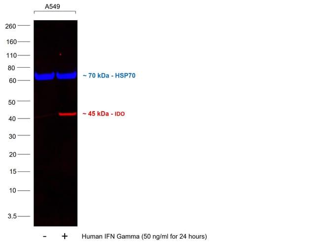
Description: This eyedio monoclonal antibody reacts with human indoleamine-2,3-dioxygenase (IDO, INDO, IDO1), an intracellular enzyme that catalyzes the degradation of tryptophan to kynurenines. IDO is expressed in a wide variety of tissues and cells, including macrophages, plasmacytoid dendritic cells, and several cell lines. IDO can be induced in many different cell types by IFN gamma or other inflammatory stimuli. Expression of IDO in antigen presenting cells and tumors is thought to mediate immune suppression through depletion of this essential amino acid and/or by the creation of tryptophan metabolites that cause apoptosis of T cells and induction of regulatory T cells. Applications Reported: This eyedio antibody has been reported for use in intracellular staining followed by flow cytometric analysis. Applications Tested: This eyedio antibody has been pre-titrated and tested by intracellular staining and flow cytometric analysis of stimulated normal human peripheral blood cells using the Intracellular Fixation & Permeabilization Buffer Set (cat. 88-8824) and protocol. Please refer to Best Protocols: Protocol A: Two step protocol for (cytoplasmic) intracellular proteins located under the Resources Tab online. This antibody can be used at 5 μL (0.06 μg) per test. A test is defined as the amount (μg) of antibody that will stain a cell sample in a final volume of 100 μL.
Specifications
Specifications
| Antigen | IDO |
| Applications | Flow Cytometry, Western Blot |
| Classification | Monoclonal |
| Clone | eyedio |
| Concentration | 5 μL/Test |
| Conjugate | eFluor 660 |
| Formulation | PBS with BSA and 0.09% sodium azide; pH 7.2 |
| Gene | Ido1 |
| Gene Accession No. | P14902 |
| Gene Alias | EC 1.13.11.52; IDO; IDO1; IDO-1; INDO; indolamine 2,3 dioxygenase; indole 2,3-dioxygenase; indoleamine; indoleamine 2,3-dioxygenase 1; indoleamine 23-dioxygenase; Indoleamine-2; indoleamine-pyrrole 2,3 dioxygenase; indoleamine-pyrrole 2,3-dioxygenase |
| Show More |
Frequently Asked Questions (FAQs)
Our options will depend on the samples you are analyzing.
If cell viability is not critical, you can store your stained samples at 4 degrees C or on ice overnight in the dark and analyze the following day.
For samples stained with eFluor organic fluorochromes, we recommend that cells be suspended in 100 uL of Flow Cytometry Staining Buffer (Cat. No. 00-4222) and 100 uL of eBioscience IC Fixation Buffer (Cat. No. 00-8222); samples can be incubated for up to 3 days at 4 degrees C in the dark. Alternatively, the 1-step Fix/Lyse Solution (Cat. No. 00-5333) can be used. This is a great option when working with whole blood but also works for other cell types.
Yes, the eFluor Organic fluorochromes can be used for intracellular staining. The eFluor organic fluorochromes maintain bright signal and require minimal changes in compensation when fixed with eBioscience IC Fixation Buffer (Cat. No. 00-8222-49) and Permeabilization Buffer (Cat. No. 00-8333-56) or 1-step Fix/Lyse Solution (Cat. No. 00-5333-54, 00-5333-57) (as compared to live cells).
Yes, in-house studies have demonstrated that the eFluor 660 fluorochrome is recognized by Anti-Cy5/Alexa Fluor 647 beads. Side by side studies with Alexa Fluor 647 versus eFluor 660 conjugated antibodies have demonstrated comparable results.
The eFluor Organic fluorochromes and eVolve QDots can be used with flow staining buffers containing PBS and protein.
The eFluor Organic Dyes (eFluor 450, APC-eFluor 780, PerCP-eFluor 710, eFluor 710) are conventional fluorochromes. In contrast, the eVolve line of products are Quantum dots.
Safety and Handling
For Research Use Only.
By clicking Submit, you acknowledge that you may be contacted by Fisher Scientific in regards to the feedback you have provided in this form. We will not share your information for any other purposes. All contact information provided shall also be maintained in accordance with our Privacy Policy.