Learn More
Invitrogen™ IL-9 Monoclonal Antibody (MH9D1), eFluor™ 450, eBioscience™


Description
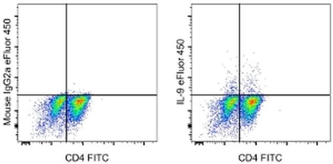
The monoclonal antibody MH9D1 recognizes interleukin-9 (IL-9), a proinflammatory cytokine historically believed to be involved in type 2 immune responses. Recent evidence suggests IL-9 may instead be secreted by a distinct T-helper lineage called Th9. These cells can be derived from Th2 cells with TGF beta or differentiated directly from naive CD4+ T cells with TGF beta and IL-4. IL-9 is a member of the common cytokine receptor gamma chain-dependent family of cytokines which also includes IL-2, IL-4, IL-7, IL-15 and IL-21. Its pleiotropic effects on Th2 lymphocytes, B lymphocytes, mast cells, eosinophils and gut and airway epithelial cells have implicated IL-9 in asthma and other allergy-related disorders. Staining with the MH9D1 antibody is not blocked by the MH9A4 antibody, suggesting that the two antibodies bind to unique epitopes. Applications Reported: This MH9D1 antibody has been reported for use in intracellular staining followed by flow cytometric analysis. Applications Tested: This MH9D1 antibody has been pre-titrated and tested by intracellular staining and flow cytometric analysis of Th9 polarized human peripheral blood cells using the Intracellular Fixation & Permeabilization Buffer Set (Product # 88-8824-00) and protocol. Please refer to BestProtocols: Protocol A: Two step protocol for (cytoplasmic) intracellular proteins located under the Resources Tab online. This antibody can be used at 5 μL (0.125 μg) per test. A test is defined...
Specifications
Specifications
| Antigen | IL-9 |
| Applications | Flow Cytometry |
| Classification | Monoclonal |
| Clone | MH9D1 |
| Concentration | 5 μL/Test |
| Conjugate | eFluor 450 |
| Formulation | PBS with BSA and 0.09% sodium azide; pH 7.2 |
| Gene | Il9 |
| Gene Accession No. | P15248 |
| Gene Alias | cytokine P40; homolog of mouse T cell and mast cell growth factor 40; HP40; IL9; IL-9; ILN; Interleukin; interleukin 9; Interleukin9; interleukin-9; P40; p40 cytokine; p40 T-cell and mast cell growth factor; T-cell growth factor P40 |
| Show More |
Frequently Asked Questions (FAQs)
Our options will depend on the samples you are analyzing.
If cell viability is not critical, you can store your stained samples at 4 degrees C or on ice overnight in the dark and analyze the following day.
For samples stained with eFluor organic fluorochromes, we recommend that cells be suspended in 100 uL of Flow Cytometry Staining Buffer (Cat. No. 00-4222) and 100 uL of eBioscience IC Fixation Buffer (Cat. No. 00-8222); samples can be incubated for up to 3 days at 4 degrees C in the dark. Alternatively, the 1-step Fix/Lyse Solution (Cat. No. 00-5333) can be used. This is a great option when working with whole blood but also works for other cell types.
Yes, the eFluor Organic fluorochromes can be used for intracellular staining. The eFluor organic fluorochromes maintain bright signal and require minimal changes in compensation when fixed with eBioscience IC Fixation Buffer (Cat. No. 00-8222-49) and Permeabilization Buffer (Cat. No. 00-8333-56) or 1-step Fix/Lyse Solution (Cat. No. 00-5333-54, 00-5333-57) (as compared to live cells).
The eFluor Organic fluorochromes and eVolve QDots can be used with flow staining buffers containing PBS and protein.
The eFluor Organic Dyes (eFluor 450, APC-eFluor 780, PerCP-eFluor 710, eFluor 710) are conventional fluorochromes. In contrast, the eVolve line of products are Quantum dots.
As with other fluorochromes, we recommend minimal exposure to light to maintain optimal signal.
By clicking Submit, you acknowledge that you may be contacted by Fisher Scientific in regards to the feedback you have provided in this form. We will not share your information for any other purposes. All contact information provided shall also be maintained in accordance with our Privacy Policy.