Promotional price valid on web orders only. Your contract pricing may differ. Interested in signing up for a dedicated account number?
Learn More
Learn More


Description
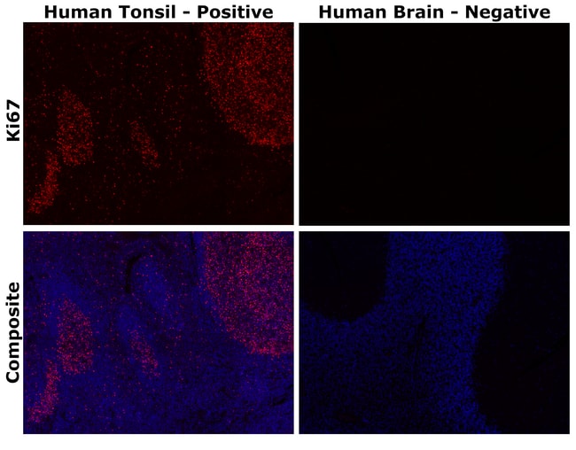
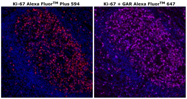
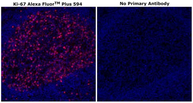
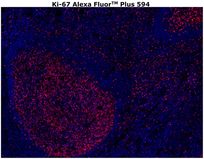
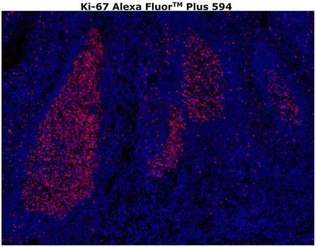
Description: The monoclonal antibody SolA15 recognizes human Ki-67, a 300 kDa nuclear protein. Ki-67 is present during all active phases of the cell cycle (G1, S, G2, and mitosis), but is absent from resting cells (G0). Ki-67 is detected within the nucleus during interphase but redistributes to the chromosomes during mitosis. Ki-67 is used as a marker for determining the growth fraction of a given population of cells. In studies of tumor cells, the 'Ki-67 labeling index' refers to the number of Ki-67 positive cells within the population and this is used to predict outcome of particular cancer types. Ki-67 has been shown to interact with the DNA-bound protein chromobox protein homolog 3 (CBX3) (heterochromatin). The SolA15 antibody also recognizes mouse, rat, non-human primate and canine Ki-67. Applications Reported: This SolA15 antibody has been reported for use in immunohistochemical staining of formalin-fixed paraffin embedded (FFPE) tissue sections and frozen (paraformaldehyde fixed) sections and immunocytochemistry (ICC). Applications Tested: This SolA15 antibody has been tested by immunohistochemistry on formalin-fixed paraffin embedded human tissue with high pH antigen retrieval at 20 μg/mL. It is recommended that this antibody be carefully titrated for optimal performance in the assay of interest. Using conjugate solutions: Centrifuge the protein conjugate solution briefly in a microcentrifuge before use; add only the supernatant to the experiment. This step w...
Ki-67 is a nuclear protein that is expressed during various stages in the cell cycle, particularly during late G1, S, G2, and M phases. The protein has a forkhead associated domain (FHA) through which it associates with euchromatin at the perichromosomal layer, the centromeric heterochromatin, and the nucleolus. Ki-67 is shown to have a cell cycle dependent topographical distribution with perinucleolar expression at G1, expression in the nuclear matrix at G2, and expression on the chromosomes during M phase. Ki-67 is commonly used as a proliferation marker because it is not detected in G0 cells, but increases steadily from G1 through mitosis. Ki-67 antibodies are useful in establishing the cell growing fraction in neoplasms. In neoplastic tissues, the prognostic value is comparable to the tritiated thymidine-labelling index. The correlation between low Ki-67 index and histologically low-grade tumors is strong. Ki-67 is routinely used as a neuronal marker of cell cycling and proliferation.Specifications
| Ki-67 | |
| Monoclonal | |
| 0.2 mg/mL | |
| PBS with 0.5% BSA, 10% proprietary stabilizer and 0.05% sodium azide; pH 7.2 | |
| P46013 | |
| Mki67 | |
| Affinity chromatography | |
| RUO | |
| 4288 | |
| 4°C, store in dark | |
| Liquid |
| Immunohistochemistry (Paraffin) | |
| SolA15 | |
| Alexa Fluor Plus 594 | |
| Mki67 | |
| antigen identified by monoclonal antibody Ki 67; antigen identified by monoclonal antibody Ki-67; Antigen identified by monoclonal antibody Ki-67 homolog; Antigen KI-67; Antigen KI-67 homolog; antigen KI-67; proliferation marker protein Ki-67; antigen KI-67-like; cb31; D630048A14Rik; I79_022666; Ki67; Ki-67; KIA; LOW QUALITY PROTEIN: proliferation marker protein Ki-67; marker of proliferation Ki-67; MIB-; MIB-1; Mki67; PPP1R105; Proliferation marker protein Ki-67; proliferation-related Ki-67 antigen; protein phosphatase 1, regulatory subunit 105; RP11-380J17.2; sb:cb31; si:ch211-250b22.7; unnamed protein product; wu:fa11g09; wu:fb57a07; wu:fi14e05 | |
| Rat | |
| 500 μL | |
| Primary | |
| Human | |
| Antibody | |
| IgG2a κ |
Product Content Correction
The Fisher Scientific Encompass Program offers items which are not part of our distribution portfolio. These products typically do not have pictures or detailed descriptions. However, we are committed to improving your shopping experience. Please use the form below to provide feedback related to the content on this product.
Product Title
Spot an opportunity for improvement?Share a Content Correction