Learn More

Invitrogen™ Ki-67 Monoclonal Antibody (SolA15), eFluor™ 570, eBioscience™, Invitrogen™


Description
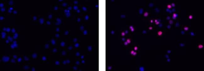
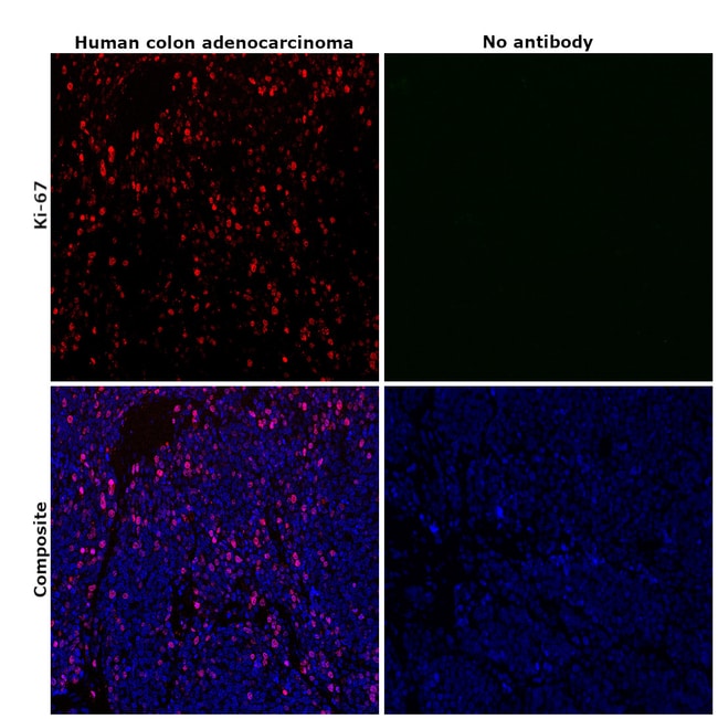
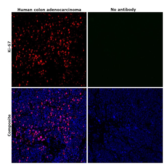
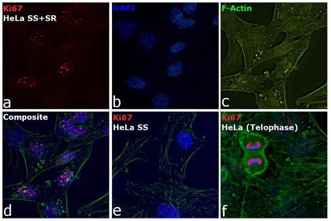
Description: The monoclonal antibody SolA15 recognizes mouse and rat Ki-67, a 300 kDa nuclear protein. Ki-67 is present during all active phases of the cell cycle (G1, S, G2, and mitosis), but is absent from resting cells (G0). Ki-67 is detected within the nucleus during interphase but redistributes to the chromosomes during mitosis. Ki-67 is used as a marker for determining the growth fraction of a given population of cells. In studies of tumor cells, the Ki-67 labeling index refers to the number of Ki-67 positive cells within the population and this is used to predict outcome of particular cancer types. Ki-67 has been shown to interact with the DNA-bound protein chromobox protein homolog 3 (CBX3) (heterochromatin). The SolA15 antibody also recognizes human, non-human primate and canine Ki-67. Applications Reported: This SolA15 antibody has been reported for use in immunohistochemical staining of frozen tissue sections, immunohistochemical staining of formalin-fixed paraffin embedded tissue sections, and immunocytochemistry. Applications Tested: This SolA15 antibody has been tested immunocytochemistry on fixed and permeabilized C2C12 cells and can be used at less than or equal to 1 μg/mL. It is recommended that the antibody be carefully titrated for optimal performance in the assay of interest.
Specifications
Specifications
| Antigen | Ki-67 |
| Applications | Immunohistochemistry (Frozen), Immunohistochemistry (Paraffin), Immunocytochemistry |
| Classification | Monoclonal |
| Clone | SolA15 |
| Concentration | 0.2 mg/mL |
| Conjugate | eFluor 570 |
| Formulation | PBS with 0.09% sodium azide; pH 7.2 |
| Gene | Mki67 |
| Gene Accession No. | E9PVX6, P46013 |
| Gene Alias | antigen identified by monoclonal antibody Ki 67; antigen identified by monoclonal antibody Ki-67; Antigen identified by monoclonal antibody Ki-67 homolog; Antigen KI-67; Antigen KI-67 homolog; antigen KI-67; proliferation marker protein Ki-67; antigen KI-67-like; cb31; D630048A14Rik; I79_022666; Ki67; Ki-67; KIA; LOW QUALITY PROTEIN: proliferation marker protein Ki-67; marker of proliferation Ki-67; MIB-; MIB-1; Mki67; PPP1R105; Proliferation marker protein Ki-67; proliferation-related Ki-67 antigen; protein phosphatase 1, regulatory subunit 105; RP11-380J17.2; sb:cb31; si:ch211-250b22.7; unnamed protein product; wu:fa11g09; wu:fb57a07; wu:fi14e05 |
| Show More |
Frequently Asked Questions (FAQs)
Our options will depend on the samples you are analyzing.
If cell viability is not critical, you can store your stained samples at 4 degrees C or on ice overnight in the dark and analyze the following day.
For samples stained with eFluor organic fluorochromes, we recommend that cells be suspended in 100 uL of Flow Cytometry Staining Buffer (Cat. No. 00-4222) and 100 uL of eBioscience IC Fixation Buffer (Cat. No. 00-8222); samples can be incubated for up to 3 days at 4 degrees C in the dark. Alternatively, the 1-step Fix/Lyse Solution (Cat. No. 00-5333) can be used. This is a great option when working with whole blood but also works for other cell types.
The eFluor Organic fluorochromes and eVolve QDots can be used with flow staining buffers containing PBS and protein.
The eFluor Organic Dyes (eFluor 450, APC-eFluor 780, PerCP-eFluor 710, eFluor 710) are conventional fluorochromes. In contrast, the eVolve line of products are Quantum dots.
As with other fluorochromes, we recommend minimal exposure to light to maintain optimal signal.
For Research Use Only.
By clicking Submit, you acknowledge that you may be contacted by Fisher Scientific in regards to the feedback you have provided in this form. We will not share your information for any other purposes. All contact information provided shall also be maintained in accordance with our Privacy Policy.