Learn More


Invitrogen™ Ki-67 Monoclonal Antibody (SolA15), eFluor™ 660, eBioscience™, Invitrogen™


Description
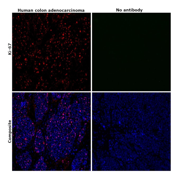
Description: The monoclonal antibody SolA15 recognizes mouse and rat Ki-67, a 300 kDa nuclear protein. Ki-67 is present during all active phases of the cell cycle (G1, S, G2, and mitosis), but is absent from resting cells (G0). Ki-67 is detected within the nucleus during interphase but redistributes to the chromosomes during mitosis. Ki-67 is used as a marker for determining the growth fraction of a given population of cells. In studies of tumor cells, the Ki-67 labeling index refers to the number of Ki-67 positive cells within the population and this is used to predict outcome of particular cancer types. Ki-67 has been shown to interact with the DNA-bound protein chromobox protein homolog 3 (CBX3) (heterochromatin). The SolA15 antibody also recognizes human, non-human primate and canine Ki-67. Applications Reported: This SolA15 antibody has been reported for use in intracellular staining followed by flow cytometric analysis, immunohistochemical staining, and immunocytochemistry. Applications Tested: This SolA15 antibody has been tested by intracellular staining and flow cytometric analysis of stimulated mouse splenocytes using the Foxp3/Transcription Factor Buffer Set (cat. 00-5523) and protocol. This can be used at less than or equal to 0.125 μg per test. A test is defined as the amount (μg) of antibody that will stain a cell sample in a final volume of 100 μL.
Specifications
Specifications
| Antigen | Ki-67 |
| Applications | Flow Cytometry, Immunohistochemistry, Immunohistochemistry (Paraffin), Immunocytochemistry |
| Classification | Monoclonal |
| Clone | SolA15 |
| Concentration | 0.2 mg/mL |
| Conjugate | eFluor 660 |
| Formulation | PBS with 0.09% sodium azide; pH 7.2 |
| Gene | Mki67 |
| Gene Accession No. | E9PVX6, P46013 |
| Gene Alias | antigen identified by monoclonal antibody Ki 67; antigen identified by monoclonal antibody Ki-67; Antigen identified by monoclonal antibody Ki-67 homolog; Antigen KI-67; Antigen KI-67 homolog; antigen KI-67; proliferation marker protein Ki-67; antigen KI-67-like; cb31; D630048A14Rik; I79_022666; Ki67; Ki-67; KIA; LOW QUALITY PROTEIN: proliferation marker protein Ki-67; marker of proliferation Ki-67; MIB-; MIB-1; Mki67; PPP1R105; Proliferation marker protein Ki-67; proliferation-related Ki-67 antigen; protein phosphatase 1, regulatory subunit 105; RP11-380J17.2; sb:cb31; si:ch211-250b22.7; unnamed protein product; wu:fa11g09; wu:fb57a07; wu:fi14e05 |
| Show More |
Frequently Asked Questions (FAQs)
Yes, the eFluor Organic fluorochromes can be used for intracellular staining. The eFluor organic fluorochromes maintain bright signal and require minimal changes in compensation when fixed with eBioscience IC Fixation Buffer (Cat. No. 00-8222-49) and Permeabilization Buffer (Cat. No. 00-8333-56) or 1-step Fix/Lyse Solution (Cat. No. 00-5333-54, 00-5333-57) (as compared to live cells).
Yes, in-house studies have demonstrated that the eFluor 660 fluorochrome is recognized by Anti-Cy5/Alexa Fluor 647 beads. Side by side studies with Alexa Fluor 647 versus eFluor 660 conjugated antibodies have demonstrated comparable results.
As with other fluorochromes, we recommend minimal exposure to light to maintain optimal signal.
For Research Use Only.
By clicking Submit, you acknowledge that you may be contacted by Fisher Scientific in regards to the feedback you have provided in this form. We will not share your information for any other purposes. All contact information provided shall also be maintained in accordance with our Privacy Policy.