Promotional price valid on web orders only. Your contract pricing may differ. Interested in signing up for a dedicated account number?
Learn More
Learn More
LIGHT Polyclonal Antibody, Biotin, PeproTech®, Invitrogen™
Rabbit Polyclonal Antibody
$250.00 - $2175.00
Specifications
| Antigen | LIGHT |
|---|---|
| Concentration | 0.1-1.0 mg/mL |
| Applications | ELISA, Western Blot |
| Classification | Polyclonal |
| Conjugate | Biotin |
| Catalog Number | Mfr. No. | Quantity | Price | Quantity & Availability | |||||
|---|---|---|---|---|---|---|---|---|---|
| Catalog Number | Mfr. No. | Quantity | Price | Quantity & Availability | |||||
500P308BT25

|
Invitrogen™
500P308BT25UG |
25 μg |
Each for $250.00
|
|
|||||
|
50-271-8524
|
Invitrogen™
500P308BT500UG |
500 μg |
Each for $2,175.00
|
|
|||||
Description
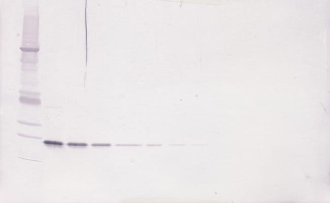
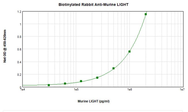
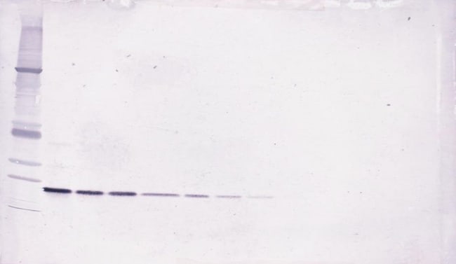
AA Sequence of recombinant protein: MRLHQRLGDI VAHLPDGGKG SWEKLIQDQR SHQANPAAHL TGANASLIGI GGPLLWETRL GLAFLRGLTY HDGALVTMEP GYYYVYSKVQ LSGVGCPQGL ANGLPITHGL YKRTSRYPKE LELLVSRRSP CGRANSSRVW WDSSFLGGVV HLEAGEEVVV RVPGNRLVRP RDGTRSYFGA FMV. Preparation: Produced from sera of rabbits immunized with highly pure Recombinant Murine LIGHT. Anti-Murine LIGHT-specific antibody was purified by affinity chromatography and then biotinylated. Sandwich ELISA: To detect Murine LIGHT by sandwich ELISA (using 100 μL/well) a concentration of 0.25-1.0 μg/mL of this antibody is required. This biotinylated polyclonal antibody, in conjunction with PeproTech Polyclonal Anti-Murine LIGHT (500-P308) as a capture antibody, allows the detection of at least 2000-4000 pg/mL of Recombinant Murine LIGHT. Western Blot: To detect Murine LIGHT by Western Blot analysis this antibody can be used at a concentration of 0.1-0.2 μg/mL. When used in conjunction with compatible secondary reagents the detection limit for Recombinant Murine LIGHT is 1.5-3.0 ng/lane, under either reducing or non-reducing conditions. 500-P308BT-1MG will be provided as 2 x 500 μg
Apoptosis, or programmed cell death, occurs during normal cellular differentiation and development of multicellular organisms. Apoptosis is induced by certain cytokines including TNF (tumor necrosis factor) and Fas ligand of the TNF family through their death domain containing receptors, TNFR1 (tumor necrosis factor receptor 1) and Fas. Cell death signals are transduced by death domain (DD)-containing adapter molecules and members of the ICE (interleukin-1-beta converting enzyme)/CED-3 (C. elegans death receptor-3) protease family. A novel DD-containing molecule was recently cloned from mouse, human and monkey and designated Daxx (death associated protein 6). Daxx binds specifically to the Fas death domain and enhances Fas induced apoptosis and activates the Jun N-terminal kinase (JNK) pathway. Daxx is widely expressed in fetal and adult human and mouse tissues indicating its important function in Fas signaling pathways.Specifications
| LIGHT | |
| ELISA, Western Blot | |
| Biotin | |
| Rabbit | |
| Mouse | |
| Q9QYH9 | |
| 50930 | |
| E.coli-derived Recombinant Murine LIGHT. | |
| Antigen affinity chromatography | |
| TNFSF14 |
| 0.1-1.0 mg/mL | |
| Polyclonal | |
| Lyophilized | |
| RUO | |
| PBS with no preservative | |
| CD258; Herpes virus entry mediator ligand; herpesvirus entry mediator ligand; HVEML; HVEM-L; Light; LTg; Ly113; sCD258; sLIGHT; soluble CD258; soluble LIGHT; Tnfsf14; TR2; tumor necrosis factor (ligand) superfamily, member 14; tumor necrosis factor ligand 1D; tumor necrosis factor ligand superfamily member 14; Tumor necrosis factor ligand superfamily member 14, membrane form; Tumor necrosis factor ligand superfamily member 14, soluble form; tumor necrosis factor superfamily member 14; UNQ391/PRO726 | |
| TNFSF14 | |
| Primary | |
| -20°C |
Spot an opportunity for improvement?Share a Content Correction
Product Content Correction
The Fisher Scientific Encompass Program offers items which are not part of our distribution portfolio. These products typically do not have pictures or detailed descriptions. However, we are committed to improving your shopping experience. Please use the form below to provide feedback related to the content on this product.
Product Title