Learn More
Invitrogen™ M-CSF Polyclonal Antibody, Biotin, PeproTech®, Invitrogen™
Goat Polyclonal Antibody
Supplier: Invitrogen™ 500P62GBT25UG

Description
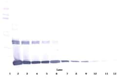
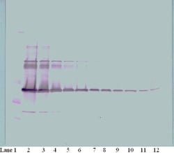
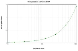
AA Sequence of recombinant protein: MKEVSEHCSH MIGNGHLKVL QQLIDSQMET SCQIAFEFVD QEQLDDPVCY LKKAFFLVQD IIDETMRFKD NTPNANATER LQELSNNLNS CFTKDYEEQN KACVRTFHET PLQLLEKIKN FFNETKNLLE KDWNIFTKNC NNSFAKCSSR DVVTKP. Preparation: Produced from sera of goats immunized with highly pure Recombinant Murine M-CSF. Anti-Murine M-CSF-specific antibody was purified by affinity chromatography and then biotinylated. Sandwich ELISA: To detect mM-CSF by sandwich ELISA (using 100 μL/well antibody solution) a concentration of 0.25-1.0 μg/mL of this antibody is required. This biotinylated polyclonal antibody, in conjunction with PeproTech Polyclonal Anti-Murine M-CSF (500-P62G) as a capture antibody, allows the detection of at least 0.2-0.4 ng/well of Recombinant mM-CSF. Western Blot: To detect mM-CSF by Western Blot analysis this antibody can be used at a concentration of 0.1-0.2 μg/mL. Used in conjunction with compatible secondary reagents the detection limit for Recombinant mM-CSF is 1.5-3.0 ng/lane, under either reducing or non-reducing conditions. 500-P62GBT-1MG will be provided as 2 x 500 μg
M-CSF (Macrophage colony-stimulating factor, CSF-1) is a survival factor essential for the proliferation and development of monocytes, macrophages, and osteoclast progenitor cells. M-CSF also induces VEGF (vascular endothelial growth factor) secretion by macrophages, thereby mediating mobilization of endothelial progenitor cells and neovascularization. M-CSF is present as several bioactive isoforms that differ in potency and stability. The full-length protein is synthesized as a membrane-spanning protein that can be expressed on the cell surface or further cleaved and modified in the secretory vesicle. Further, M-CSF is a disulfide-bonded homodimer which is processed into one of two isoforms, a glycoprotein or a proteoglycan that has been modified by the addition of chondroitin sulfate to each subunit. Binding of M-CSF to its receptor, c-Fms (CSF-1R or CD115) induces dimerization of the receptor followed by internalization and degradation of the complex. Functionally, M-CSF is known to stimulate differentiation of hematopoietic stem cells to monocyte-macrophage cell populations in culture. M-CSF acts through the CSF receptor 1. Although human M-CSF shows activity on mouse cells, mouse CSF shows no activity on human cells.
Specifications
| M-CSF | |
| Polyclonal | |
| Biotin | |
| Csf1 | |
| C87615; colony stimulating factor 1; colony stimulating factor 1 (macrophage); colony-stimulating factor-1 splice variant; CSF1; CSF-1; Csfm; H-MCSF; lanimostim; macrophage colony stimulating factor 1; macrophage colony stimulating factor alpha; macrophage colony stimulating factor beta; macrophage colony-stimulating factor; macrophage colony-stimulating factor 1; macrophage colony-stimulating factor beta; macrophage-colony stimulating factor alpha; MCSF; MCSF alpha; MCSFBETA; MGC31930; M-MCSF; op; osteopetrosis; Processed macrophage colony-stimulating factor 1 | |
| Goat | |
| Antigen Affinity Chromatography | |
| RUO | |
| 12977 | |
| -20°C | |
| Lyophilized |
| ELISA, Western Blot | |
| 0.1-1.0 mg/mL | |
| PBS with no preservative | |
| P07141 | |
| Csf1 | |
| E.coli-derived Recombinant Murine M-CSF. | |
| 25 μg | |
| Primary | |
| Mouse | |
| Antibody |
The Fisher Scientific Encompass Program offers items which are not part of our distribution portfolio. These products typically do not have pictures or detailed descriptions. However, we are committed to improving your shopping experience. Please use the form below to provide feedback related to the content on this product.


