Promotional price valid on web orders only. Your contract pricing may differ. Interested in signing up for a dedicated account number?
Learn More
Learn More
Description
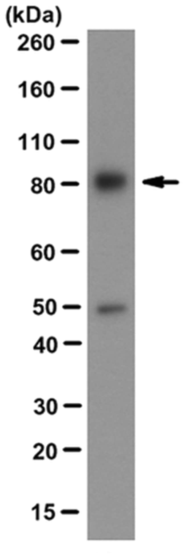
Specifically detects XPB clone: 15TF2-1B3 in Human samples, and it is validated for Immunocytochemistry, Western Blotting
Transcription factor II human (TFIIH) basal transcription factor complex helicase XPB subunit (EC 3.6.4.12; UniProt P19447; also known as BTF2 p89, DNA excision repair protein ERCC-3, DNA repair helicase, DNA repair protein complementing XP-B cells, TFIIH 89kDa subunit, TFIIH basal transcription factor complex 89kDa subunit, TFIIH p89, Basic transcription factor 2 89kDa subunit, Xeroderma pigmentosum group B-complementing protein) is encoded by the ERCC3 (also known as BTF2, GTF2H, RAD25, TFIIH, XPB) gene (Gene ID 2071) in human. DNA lesions caused by UV irradiation, drugs, or other environmental factors are eliminated by two nucleotide excision repair (NER) pathways, Global genome repair (GGR) and transcription-coupled repair (TCR). In GGR, the removal of lesions requires their recognition by the repair factor XPC/HR23b and the subsequent opening of the DNA duplex by TFIIH. The resulting single-stranded structure is stabilized by XPA and replication protein A (RPA). XPG is recruited through its interaction with TFIIH on the 3′ side of the lesion and its positioning on the cut site requires RPA. The interaction between XPA and XPB (ERCC1) stimulates the recruitment of ERCC1-XPF on the 5′ side of the DNA lesion. The damaged oligonucleotide can then be removed through the double incision by XPG and ERCC1-XPF endonucleases. In TCR, these factors (except XPC/HR23B) are recruited by the stalled RNA pol II in front of the damage with the help of the CSB and CSA proteins.
Specifications
Specifications
| Antigen | XPB |
| Applications | Immunocytochemistry, Western Blot |
| Classification | Monoclonal |
| Clone | 15TF2-1B3 |
| Formulation | Mouse monoclonal IgG1κ ascites with 0.05% sodium azide. |
| Gene Accession No. | P19447 |
| Gene Symbols | ERCC3; XPB; XPBC |
| Host Species | Mouse |
| Immunogen | Recombinant protein corresponding to human XPB. |
| Purification Method | Unpurified |
| Show More |
For Research Use Only
Product Title
By clicking Submit, you acknowledge that you may be contacted by Fisher Scientific in regards to the feedback you have provided in this form. We will not share your information for any other purposes. All contact information provided shall also be maintained in accordance with our Privacy Policy.
Spot an opportunity for improvement?