Learn More
Invitrogen™ Myeloperoxidase (MPO) Monoclonal Antibody (MPO455-8E6), eFluor™ 450, eBioscience™


Description
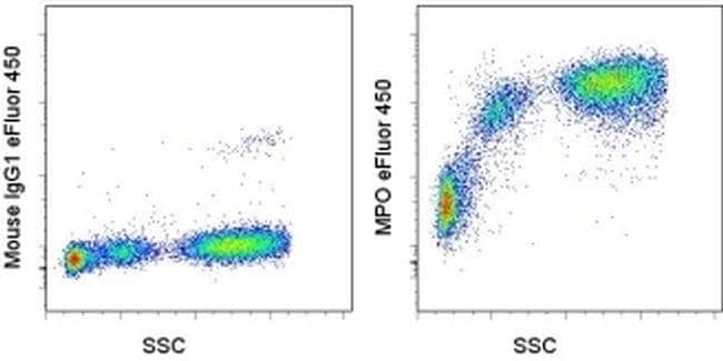
Description: The monoclonal antibody MPO455-8E6 recognizes myeloperoxidase (MPO), a protein within the azurophilic granules of myeloid cells. MPO is a multimeric protein comprised of two 55 kDa subunits and two 15 kDa subunits. The larger subunits associate with a heme protein resulting in a greenish color. As an enzyme, MPO breaks down hydrogen peroxide and oxidizes tyrosine. The products of this reaction, hypochlorous acid and tyrosyl radical, causethe cytotoxic and killing effects characteristic of neutrophils. Myeloperoxidase is important in the diagnosis of some cancers and increases in serum levels have been shown to correlate with cardiac events. Applications Reported: This MPO455-8E6 antibody has been reported for use in flow cytometric analysis. Applications Tested: This MPO455-8E6 antibody has been pre-titrated and tested by intracellular staining followed by flow cytometric analysis of normal human peripheral blood cells using the Intracellular Fixation & Permeablization Buffer Set (Product # 88-8824-00). For best results, whole blood should first be stained with antibodies to surface antigens then treated with 1X RBC lysis buffer (Product # 00-4333-μ) to lyse erythrocytes. Then, for intracellular staining for MPO, cells should be fixed with IC Fixation Buffer (Product # 00-8222-49) washed two times with Permeabilization Buffer (Product # 00-8333-56) and then incubated with MPO455-8E6 for 30-60 minutes. After washing, cells may be analyzed on a flow cytom...
Specifications
Specifications
| Antigen | Myeloperoxidase (MPO) |
| Applications | Flow Cytometry |
| Classification | Monoclonal |
| Clone | MPO455-8E6 |
| Concentration | 5 μL/Test |
| Conjugate | eFluor 450 |
| Formulation | PBS with BSA and 0.09% sodium azide; pH 7.2 |
| Gene | MPO |
| Gene Accession No. | P05164 |
| Gene Alias | 84 kDa myeloperoxidase; 89 kDa myeloperoxidase; mKIAA4033; MPO; Myeloperoxidase; Myeloperoxidase heavy chain; Myeloperoxidase light chain |
| Show More |
Frequently Asked Questions (FAQs)
Our options will depend on the samples you are analyzing.
If cell viability is not critical, you can store your stained samples at 4 degrees C or on ice overnight in the dark and analyze the following day.
For samples stained with eFluor organic fluorochromes, we recommend that cells be suspended in 100 uL of Flow Cytometry Staining Buffer (Cat. No. 00-4222) and 100 uL of eBioscience IC Fixation Buffer (Cat. No. 00-8222); samples can be incubated for up to 3 days at 4 degrees C in the dark. Alternatively, the 1-step Fix/Lyse Solution (Cat. No. 00-5333) can be used. This is a great option when working with whole blood but also works for other cell types.
Yes, the eFluor Organic fluorochromes can be used for intracellular staining. The eFluor organic fluorochromes maintain bright signal and require minimal changes in compensation when fixed with eBioscience IC Fixation Buffer (Cat. No. 00-8222-49) and Permeabilization Buffer (Cat. No. 00-8333-56) or 1-step Fix/Lyse Solution (Cat. No. 00-5333-54, 00-5333-57) (as compared to live cells).
The eFluor Organic fluorochromes and eVolve QDots can be used with flow staining buffers containing PBS and protein.
The eFluor Organic Dyes (eFluor 450, APC-eFluor 780, PerCP-eFluor 710, eFluor 710) are conventional fluorochromes. In contrast, the eVolve line of products are Quantum dots.
By clicking Submit, you acknowledge that you may be contacted by Fisher Scientific in regards to the feedback you have provided in this form. We will not share your information for any other purposes. All contact information provided shall also be maintained in accordance with our Privacy Policy.