Promotional price valid on web orders only. Your contract pricing may differ. Interested in signing up for a dedicated account number?
Learn More
Learn More
p57 Kip2 Antibody (KIP2/880), Novus Biologicals™


Mouse Monoclonal Antibody
$268.00 - $524.00
Specifications
| Antigen | p57 Kip2 |
|---|---|
| Clone | KIP2/880 |
| Concentration | 0.2mg/mL |
| Dilution | Flow Cytometry 0.5 - 1 ug/million cells in 0.1 ml, Immunohistochemistry-Paraffin 0.25 - 0.5 ug/ml, Immunofluorescence 0.5 - 1.0 ug/ml |
| Applications | Flow Cytometry, Immunohistochemistry (Paraffin), Immunofluorescence |
| Catalog Number | Mfr. No. | Quantity | Price | Quantity & Availability | |||||
|---|---|---|---|---|---|---|---|---|---|
| Catalog Number | Mfr. No. | Quantity | Price | Quantity & Availability | |||||
NBP24449000
 |
Novus Biologicals
NBP2444900.02MG |
0.02 mg |
Each for $268.00
|
|
|||||
NBP24449001
 |
Novus Biologicals
NBP2444900.1MG |
0.1 mg |
Each for $524.00
|
|
|||||
NBP24449002
 |
Novus Biologicals
NBP2444900.2MG |
0.2 mg | N/A | N/A | N/A | ||||
Description
Ensure accurate, reproducible results in Flow Cytometry, Immunohistochemistry (Paraffin), Immunofluorescence
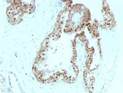
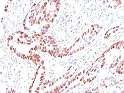
p57 Kip2 Monoclonal specifically detects p57 Kip2 in Human, Mouse samples. It is validated for Flow Cytometry, Immunohistochemistry, Immunocytochemistry/Immunofluorescence, Immunohistochemistry-Paraffin, Immunofluorescence.Specifications
| p57 Kip2 | |
| 0.2mg/mL | |
| Flow Cytometry, Immunohistochemistry (Paraffin), Immunofluorescence | |
| Unconjugated | |
| Mouse | |
| Breast Cancer, Cancer, Cell Cycle and Replication, Core ESC Like Genes, DNA Repair, Stem Cell Markers | |
| 10mM PBS and 0.05% BSA with 0.05% Sodium Azide | |
| Beckwith-Wiedemann syndrome, BWS, cyclin-dependent kinase inhibitor 1C, cyclin-dependent kinase inhibitor 1C (p57, Kip2), Cyclin-dependent kinase inhibitor p57, KIP2BWCR, p57, p57Kip2, WBS | |
| CDKN1C | |
| IgG2b κ | |
| Protein A or G purified | |
| Recognizes a protein of 57kDa, identified as p57Kip2. It shows no cross-reaction with p27Kip1. p57Kip2 is a potent tight-binding inhibitor of several G1 cyclin complexes, and is a negative regulator of cell proliferation. Anti-p57 has been used as an aide in identification of complete hydatidiform mole (CHM) (no nuclear labeling of cytotrophoblasts and stromal cells) from partial hydatidiform mole (PHM) in which both cytotrophoblasts and stromal cells stain. The histological differentiation of complete mole, partial mole, and hydropic spontaneous abortion is problematic. Most complete hydatidiform moles are diploid, whereas most partial moles are triploid. Ploidy studies will identify partial moles, but will not differentiate complete moles from non-molar gestations. Complete moles carry a high risk of persistent disease and choriocarcinoma, while partial moles have a very low risk. In normal placenta, many cytotrophoblast nuclei and stromal cells are labeled with this antibody. Similar findings apply to PHM and hydropic abortus tissues. Intervillous trophoblastic islands (IVTIs) demonstrate nuclear labeling in all three entities and serve as an internal control. |
| KIP2/880 | |
| Flow Cytometry 0.5 - 1 ug/million cells in 0.1 ml, Immunohistochemistry-Paraffin 0.25 - 0.5 ug/ml, Immunofluorescence 0.5 - 1.0 ug/ml | |
| Monoclonal | |
| Purified | |
| RUO | |
| Human, Mouse | |
| P49918 | |
| 1028 | |
| Recombinant human p57Kip2 protein | |
| Primary | |
| Store at 4C. | |
| 57 kDa |
For Research Use Only
Spot an opportunity for improvement?Share a Content Correction
Product Content Correction
Your input is important to us. Please complete this form to provide feedback related to the content on this product.
Product Title

