Learn More
Invitrogen™ PCNA Monoclonal Antibody (PC10 (3F81)), eFluor™ 615, eBioscience™


Description
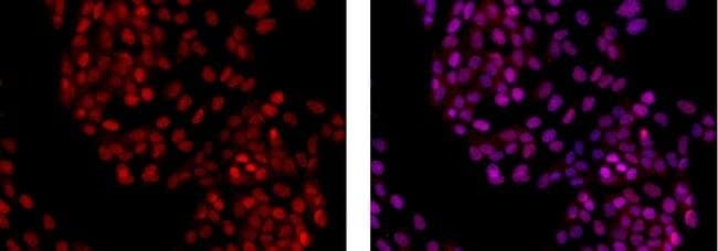
The PC10 antibody recognizes the proliferating cell nuclear antigen (PCNA), a 36 kDa protein, also known as polymerase delta auxiliary protein. PC10 antibody reacts with human, mouse, and rat PCNA. The peak expression of PCNA occurs during the S-phase. Applications Reported: This PC10 (a.k.a. 3F81) antibody has been reported for use in immunohistochemical and immunocytochemical staining. Applications Tested: This PC10 (a.k.a. 3F81) antibody has been tested by immunocytochemistry of methanol-fixed MDCK cells and can be used at less than or equal to 0.5 μg/mL. It is recommended that the antibody be carefully titrated for optimal performance in the assay of interest. This product has not been validated for flow cytometric analysis. Filter Recommendation: When using this eFluor™ 615 antibody conjugate, we recommend a filter that will capture the 615 emission wavelength (for example, Excitation 560/55, 585LP, Emission 645/75). A standard Alexa Fluor™ 594 filter is acceptable. Excitation: 595 nm; Emission: 615 nm. Filtration: 0.2 μm post-manufacturing filtered.
Specifications
Specifications
| Antigen | PCNA |
| Applications | Immunohistochemistry, Immunocytochemistry |
| Classification | Monoclonal |
| Clone | PC10 (3F81) |
| Concentration | 0.2 mg/mL |
| Conjugate | eFluor 615 |
| Formulation | PBS with 0.09% sodium azide; pH 7.2 |
| Gene | Pcna |
| Gene Accession No. | P04961, P12004, P17918 |
| Gene Alias | ATLD2; cb16; Cyclin; DNA polymerase delta auxiliary protein; etID36690.10; fa28e03; fb36g03; HGCN8729; hypothetical protein LOC515499; MGC8367; Pcna; pcna protein; Pcna/cyclin; PCNAR; POL30; Proliferating cell nuclear antigen; wu:fa28e03; wu:fb36g03; YBR0811; YBR088C |
| Show More |
Frequently Asked Questions (FAQs)
Our options will depend on the samples you are analyzing.
If cell viability is not critical, you can store your stained samples at 4 degrees C or on ice overnight in the dark and analyze the following day.
For samples stained with eFluor organic fluorochromes, we recommend that cells be suspended in 100 uL of Flow Cytometry Staining Buffer (Cat. No. 00-4222) and 100 uL of eBioscience IC Fixation Buffer (Cat. No. 00-8222); samples can be incubated for up to 3 days at 4 degrees C in the dark. Alternatively, the 1-step Fix/Lyse Solution (Cat. No. 00-5333) can be used. This is a great option when working with whole blood but also works for other cell types.
Yes, the eFluor Organic fluorochromes can be used for intracellular staining. The eFluor organic fluorochromes maintain bright signal and require minimal changes in compensation when fixed with eBioscience IC Fixation Buffer (Cat. No. 00-8222-49) and Permeabilization Buffer (Cat. No. 00-8333-56) or 1-step Fix/Lyse Solution (Cat. No. 00-5333-54, 00-5333-57) (as compared to live cells).
The eFluor Organic fluorochromes and eVolve QDots can be used with flow staining buffers containing PBS and protein.
As with other fluorochromes, we recommend minimal exposure to light to maintain optimal signal.
By clicking Submit, you acknowledge that you may be contacted by Fisher Scientific in regards to the feedback you have provided in this form. We will not share your information for any other purposes. All contact information provided shall also be maintained in accordance with our Privacy Policy.