Learn More
Invitrogen™ Perforin Monoclonal Antibody (dG9 (delta G9)), eFluor™ 450, eBioscience™, Invitrogen™


Description
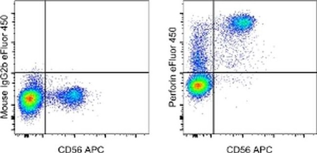
Description: The dG9 antibody clone reacts with human perforin (pore-forming protein, pfp). Perforin is one of the cytolytic mediators present in the cytoplasmic granules of cytotoxic T lymphocytes (CTL) and natural killer cells (NK). Perforin is involved in the killing function by CTLs and NKs and has an important role in the immune response against tumors and virus infections. Applications Reported: This dG9 (delta G9) antibody has been reported for use in intracellular staining followed by flow cytometric analysis. Applications Tested: This dG9 (delta G9) antibody has been pre-titrated and tested by intracellular staining followed by flow cytometric analysis of normal human peripheral blood cells using the Intracellular Fixation & Permeabilization Buffer Set (cat. 88-8824) and protocol. Please refer to Best Protocols: Protocol A: Two step protocol for (cytoplasmic) intracellular proteins located under the Resource Tab online. This can be used at 5 μL (0.125 μg) per test. A test is defined as the amount (μg) of antibody that will stain a cell sample in a final volume of 100 μL. Cell number should be determined empirically but can range from 10^5 to 10^8 cells/test. eFluor™ 450 is an alternative to Pacific Blue™. eFluor™ 450 emits at 445 nm and is excited with the Violet laser (405 nm). Please make sure that your instrument is capable of detecting this fluorochome.
Specifications
Specifications
| Antigen | Perforin |
| Applications | Flow Cytometry |
| Classification | Monoclonal |
| Clone | dG9 (delta G9) |
| Concentration | 5 μL/Test |
| Conjugate | eFluor 450 |
| Formulation | PBS with BSA and 0.09% sodium azide; pH 7.2 |
| Gene | PRF1 |
| Gene Accession No. | P14222 |
| Gene Alias | Cyta; Cytolysin; FLH2; HPLH2; lymphocyte pore forming protein; lymphocyte pore-forming protein; OMAK; OTTHUMP00000019759; P1; perforin 1; perforin 1 (pore forming protein); Perforin1; perforin-1; Pfn; PFN1; Pfp; PGFL; PIGF; PIGF-2; PLGF; pore forming protein; pore-forming protein; PRF1; Prf-1; PRF1 (pore forming protein 1); RATCYTA; RP11-710A11.3 |
| Show More |
Frequently Asked Questions (FAQs)
Our options will depend on the samples you are analyzing.
If cell viability is not critical, you can store your stained samples at 4 degrees C or on ice overnight in the dark and analyze the following day.
For samples stained with eFluor organic fluorochromes, we recommend that cells be suspended in 100 uL of Flow Cytometry Staining Buffer (Cat. No. 00-4222) and 100 uL of eBioscience IC Fixation Buffer (Cat. No. 00-8222); samples can be incubated for up to 3 days at 4 degrees C in the dark. Alternatively, the 1-step Fix/Lyse Solution (Cat. No. 00-5333) can be used. This is a great option when working with whole blood but also works for other cell types.
Yes, the eFluor Organic fluorochromes can be used for intracellular staining. The eFluor organic fluorochromes maintain bright signal and require minimal changes in compensation when fixed with eBioscience IC Fixation Buffer (Cat. No. 00-8222-49) and Permeabilization Buffer (Cat. No. 00-8333-56) or 1-step Fix/Lyse Solution (Cat. No. 00-5333-54, 00-5333-57) (as compared to live cells).
The eFluor Organic fluorochromes and eVolve QDots can be used with flow staining buffers containing PBS and protein.
The eFluor Organic Dyes (eFluor 450, APC-eFluor 780, PerCP-eFluor 710, eFluor 710) are conventional fluorochromes. In contrast, the eVolve line of products are Quantum dots.
As with other fluorochromes, we recommend minimal exposure to light to maintain optimal signal.
For Research Use Only.
By clicking Submit, you acknowledge that you may be contacted by Fisher Scientific in regards to the feedback you have provided in this form. We will not share your information for any other purposes. All contact information provided shall also be maintained in accordance with our Privacy Policy.