Promotional price valid on web orders only. Your contract pricing may differ. Interested in signing up for a dedicated account number?
Learn More

Invitrogen™ Phospho-Histone H2A.X (Ser139) Monoclonal Antibody (CR55T33), PE, eBioscience™
GREENER_CHOICE

Catalog No. 501122481
Encompass_Preferred
Change view
Click to view available options
Quantity:
100 Tests
1 product options available for selection
Product selection table with 1 available options. Use arrow keys to navigate and Enter or Space to select.
Catalog No. Quantity
50-112-2481 100 Tests
Use arrow keys to navigate between rows. Press Enter or Space to select a product option. 1 options available.
1 options
Catalog No. 50-112-2481 Supplier Invitrogen™ Supplier No. 12986542
Add to Cart
Edge
Add to Cart

Mouse Monoclonal Antibody

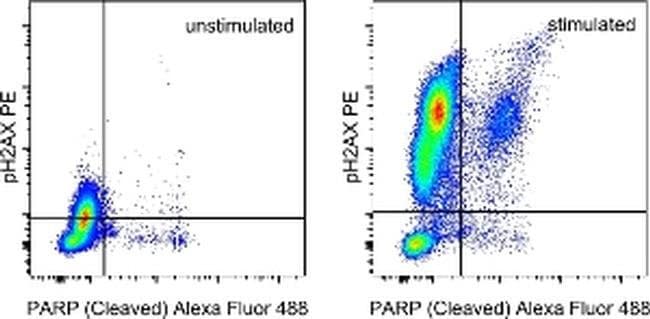
Description: The CR55T33 monoclonal antibody recognizes phosphorylated serine 139 of human and mouse H2AX. H2AX is a member of the H2A histone family that complex with DNA and other histones to form the repeating nucleosome units characteristic of eukaryotic chromatin. Nucleosomes consist of approximately 147 base pairs of DNA wrapped around an octamer of histones composed of two each of the four histone proteins: H2A, H2B, H3 and H4. After induction of DNA damage such as double-strand breaks by irradiation, genotoxic stresses, replication errors or gene recombination, PI3K-like kinases (e.g., ataxia telangiectasia mutated (ATM), ataxia telangiectasia Rad-3-related (ATR), and DNA-dependent protein kinase (DNA-PK) are activated to phosphorylate serine 139 in H2AX. This early phosphorylation event plays a critical role in recruiting proteins involved in DNA repair. The monoclonal antibody CR55T33 recognizes a single band of approximately 15 kDa on reduced cell lysates from Jurkat cells stimulated with etoposide. Applications Reported: This CR55T33 antibody has been reported for use in intracellular staining followed by flow cytometric analysis. Applications Tested: This CR55T33 antibody has been pre-titrated and tested by intracellular staining and flow cytometric analysis of stimulated normal human peripheral blood cells using the Foxp3/Transcription Factor Staining Buffer Set (Product # 00-5523-00) and protocol. Please refer to BestProtocols®: Protocol B: One step prot...

Histone H2A.X (H2AX) is a member of the histone H2A family which is one of the four core histones making up the nucleosome core particle. In eukaryotes, DNA double strand breaks (DSBs) have been shown to trigger the phosphorylation of serine 139 at the carboxy terminus of histone H2AX resulting in gamma-H2AX. The phosphorylation of H2AX can be detected by Western blotting or immunofluorescence, revealing the frequency of DSBs. The phosphatidylinositol 3-kinases have been implicated in H2AX phosphorylation, but it is unclear if ATM is the primary H2AX kinase or if other members of the family such as DNA-PK and ATR contribute in a similar manner. Structurally, H2A.x contains 143 amino acid residues. Histone H2A.X is considered a basal histone, being synthesized in G1 as well as in S-phase, and its mRNA contains polyA addition motifs and a polyA tail along with the conserved stem-loop and U7 binding sequences involved in the processing and stability of replication type histone mRNAs. There are two forms of Histone H2A.X mRNA, one about 1600 bases long and contains polyA; the other about 575 bases long, lacking polyA. The short form behaves as a replication type histone mRNA, while the longer behaves as a basal type histone mRNA. Histone H2A.X maps to the 11q23.2-q23.3 region of the human chromosome. Histone H2A.x contributes to histone-formation and therefore the structure of DNA. Histone H2A variant H2A.x specifically regulates the interaction of MDC1 (mediator of DNA damage checkpoint protein 1), a DNA repair protein to the sites of DNA damage.
TRUSTED_SUSTAINABILITY

Specifications

Antigen Phospho-Histone H2A.X (Ser139)
Applications Flow Cytometry
Classification Monoclonal
Clone CR55T33
Concentration 5 μL/Test
Conjugate PE
Formulation PBS with BSA and 0.09% sodium azide; pH 7.2
Gene H2AX
Gene Accession No. P16104, P27661
Gene Alias AW228881; gamma H2AX; gammaH2ax; H2A histone family member X; H2A histone family, member X; H2A.X; H2A.X variant histone; H2A/X; h2afx; H2ax; H2AX histone; Hist5-2ax; histone 5 protein 2ax; histone H2A.x; Histone H2a/x; Histone H2AX; RGD1566119; similar to H2A histone family, member X; zgc:56329
Gene Symbols H2AX
Host Species Mouse
Purification Method Affinity chromatography
Quantity 100 Tests
Regulatory Status RUO
Primary or Secondary Primary
Gene ID (Entrez) 15270, 3014
Target Species Human, Mouse
Content And Storage 4°C, store in dark, DO NOT FREEZE!
Product Type Antibody
Form Liquid
Isotype IgG1 κ
Show More Show Less
How does eBioscience determine the specificity of their phosphorylation-specific antibodies?

Antibodies are tested and validated in a variety of ways. First, we verify that in a panel of cells in which different pathways have been induced, we see phosphorylation specific staining only in the cells in which the specific pathway of interest has been activated. Second, we verify that phosphorylation specific staining is observed only in cell types in which the protein is expressed and not in cell types in which the protein is not expressed. Third, whenever possible, western immunoblotting is used to confirm the presence of a band(s) of the appropriate size(s) in stimulated/treated cells (and not in unstimulated/untreated cells, as appropriate).

How long can cells that have been fixed and placed in methanol be stored for use with eBioscience antibodies? What is the recommended storage temperature? How about storage of fixed cells in eBioscience fixation buffers?

We have data demonstrating that fixed cells stored in methanol are stable at -20 degrees C or at -80 degrees C for several weeks. We do not recommend storing fixed cells in eBioscience Foxp3/Transcription Factor Buffer. However, cells fixed with eBioscience Foxp3/Transcription Factor Buffer or eBioscience Intracellular (IC) fixation buffer can be stored in eBioscience IC fixation buffer for up to 3 days at 4 degrees C in the dark.
Note: Please be aware that higher compensation values may be seen with tandem dyes if any other fixative is used apart from eBioscience IC fixation buffer, for 3-day storage.

What is the optimal protocol for eBioscience phosphorylation-specific antibodies?

Each antibody has been tested in three different buffer systems: IC fixation and permeabilization, Foxp3/Transcription Factor Buffer, and IC fixation Buffer/Methanol. The recommended buffer system(s) will be noted on the Technical Data Sheet for the specific antibody.

What is the best way to maintain surface staining when using the methanol-based protocol?

Because methanol can destroy the epitope recognized by some antibodies, ideally surface staining would be performed at the beginning of the protocol with a methanol-resistant fluorochrome (like eFluor 450, FITC, and eFluor 660).
Please recognize that surface staining before stimulation can have undesired effects due to activation of the cell caused by antibody binding. We recommend staining after stimulation/treatment, fixation, and methanol treatment.

Can I use other buffer systems from other companies when using eBioscience phosphorylation-specific antibodies?

In general, you can use other companies' buffer systems provided that their buffers are of a similar composition as the buffers recommended. Be aware that results will vary depending upon the buffer system used. eBioscience antibodies have been optimized for use in eBioscience buffers and we have not tested all of our antibodies in other buffer systems. Thus, we highly recommend using our optimized protocol and buffer systems.

What are the appropriate controls to run when using eBioscience phosphorylation-specific antibodies?

It is important to understand whether the stimulation/treatment results in an upregulation or a down regulation of the phosphorylation event. Thus, it is recommended that changes in phosphorylation levels in stimulated/treated versus unstimulated/untreated cells be compared. A histogram or density plot overlay these two treatments will provide a better assessment of a change in phosphorylation event than using isotype controls.

Can I perform phosphorylation specific staining and stain for intracellular cytokines at the same time when using eBioscience phosphorylation-specific antibodies?

In theory, if the buffer systems are compatible for the antibodies of interest, this can be done. In practice, cytokine translation and phosphorylation events do not typically occur in the same timeframe, therefore, it may be difficult to detect both events in the same sample. The results will be depended on the kinetics and should be determined for each system and protein of interest.

When using eBioscience phosphorylation-specific antibodies, can I perform phosphorylation specific staining together with transcription factor staining at the same time? If so, what buffer and protocol do I use?

In theory, this can be done if the antibodies against both the transcription factor and the phosphorylated protein work in the same buffer system. For example, Anti-Human/Mouse phsopho-H2AX and all transcription factor eBioscience antibodies offered will work in the Foxp3/Transcription Factor Buffer System (Cat. No. 00-5523-00).

For eBioscience phosphorylation-specific antibodies, can total and phosphorylated protein be analyzed in parallel?

In theory, this can be done if the antibodies to both proteins work in the same buffer system and if the antibodies recognize different epitopes. Please refer to the individual Technical Data Sheets for information about buffer compatibility. Also, consider using eBioscience InstantOne ELISA Kits for immunoassay quantitation of both total and phosphorylated proteins such as AKT1/2/3, JNK1/2/3, NFκB, STAT3, and STAT5.


WARNING: Reproductive Harm - www.P65Warnings.ca.gov
Product Title
Select an issue

By clicking Submit, you acknowledge that you may be contacted by Fisher Scientific in regards to the feedback you have provided in this form. We will not share your information for any other purposes. All contact information provided shall also be maintained in accordance with our Privacy Policy.