Promotional price valid on web orders only. Your contract pricing may differ. Interested in signing up for a dedicated account number?
Learn More
Learn More
Invitrogen™ Phospho-Lck (Tyr505) Recombinant Rabbit Monoclonal Antibody (LckY505-A3)
Rabbit Recombinant Monoclonal Antibody
$408.50 - $625.00
Specifications
| Antigen | Phospho-Lck (Tyr505) |
|---|---|
| Clone | LckY505-A3 |
| Concentration | 0.5 mg/mL |
| Content And Storage | -20°C |
| Applications | Flow Cytometry, Western Blot |
| Catalog Number | Mfr. No. | Quantity | Price | Quantity & Availability | |||||
|---|---|---|---|---|---|---|---|---|---|
| Catalog Number | Mfr. No. | Quantity | Price | Quantity & Availability | |||||
|
PIMA528017
|
Invitrogen™
MA528017 |
200 μL |
Each for $625.00
|
|
|||||
|
PIMA537092
|
Invitrogen™
MA537092 |
20 μL |
Each for $408.50
|
|
|||||
Description
Recombinant rabbit monoclonal antibodies are produced using in vitro expression systems. The expression systems are developed by cloning in the specific antibody DNA sequences from immunoreactive rabbits. Then, individual clones are screened to select the best candidates for production. The advantages of using recombinant rabbit monoclonal antibodies include: better specificity and sensitivity, lot-to-lot consistency, animal origin-free formulations, and broader immunoreactivity to diverse targets due to larger rabbit immune repertoire.
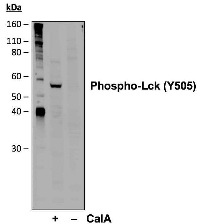
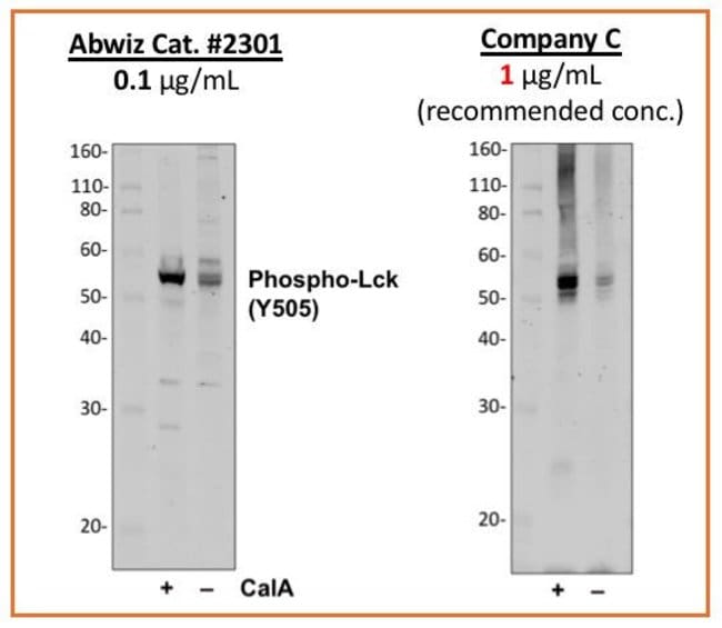
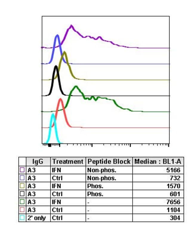
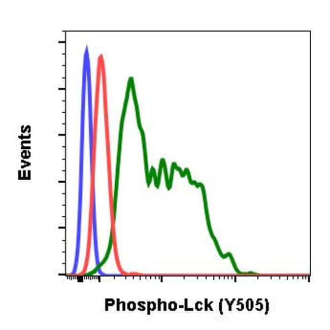
LCK is a member of the Src-family tyrosine kinase, which are essential for signaling through the T-cell receptor (TCR) complex. Upon TCR triggering, LCK phosphorylates the ITAM motives in its zeta subunits establishing binding sites for the SH2 domains of the tyrosine kinase ZAP70, which is also phosphorylated by LCK and activated to generate subsequent signaling platforms by phosphorylation of adaptor LAT. The majority of LCK is localized to the plasma membrane, however, there is also a significant fraction associated with the Golgi apparatus which may contribute to Raf activation under conditions of weak stimulation through the TCR. LCK contains N-terminal sites for myristylation and palmitylation, a PTK domain, and SH2 and SH3 domains which are involved in mediating protein-protein interactions with phosphotyrosine-containing and proline-rich motifs, respectively. LCK is also involved in the regulation of apoptosis induced by various stimuli, but not by the death receptors. Diseases associated with LCK include Immunodeficiency 22 and CD45 deficiency. Alternatively splice variants of the LCK gene encoding different isoforms of the protein have been found.Specifications
| Phospho-Lck (Tyr505) | |
| 0.5 mg/mL | |
| Flow Cytometry, Western Blot | |
| Unconjugated | |
| Rabbit | |
| RUO | |
| PBS with 0.1% BSA, 50% glycerol and 0.02% sodium azide; pH 7.4 | |
| P06239 | |
| 3932 | |
| A synthetic phospho-peptide corresponding to residues surrounding Tyr505 of human phospho Lck. | |
| Antibody |
| LckY505-A3 | |
| -20°C | |
| Recombinant Monoclonal | |
| Liquid | |
| IgG κ | |
| Human | |
| LCK | |
| EC 2.7.10.2; Hck-3; IMD22; kinase Lck; Lck; LCK proto-oncogene, Src family tyrosine kinase; Lck1; Lcktkr; leukocyte C-terminal Src kinase; LSK; Lskt; Lsk-t; Lymphocyte cell-specific protein-tyrosine kinase; lymphocyte protein tyrosine kinase; lymphocyte-specific protein tyrosine kinase; OTTHUMP00000008640; OTTHUMP00000008740; OTTHUMP00000008741; p56(LSTRA) protein-tyrosine kinase; p56>lck>; p56lck; p56-LCK; pp58lck; Protein YT16; Proto-oncogene Lck; Proto-oncogene tyrosine-protein kinase LCK; RP4-675E8.4; t cell-specific protein-tyrosine kinase; T-lymphocyte specific protein tyrosine kinase p56lck; tyr; tyrosine-protein kinase Lck; YT16 | |
| LCK | |
| Primary | |
| Protein A/G |
Spot an opportunity for improvement?Share a Content Correction
Product Content Correction
Your input is important to us. Please complete this form to provide feedback related to the content on this product.
Product Title