Learn More
Invitrogen™ Phospho-MCL-1 (Ser159) Monoclonal Antibody (RBCERNR), PE, eBioscience™


Description
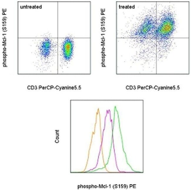
This RBCERNR monoclonal antibody recognizes human and mouse myeloid cell leukemia sequence 1 (Mcl-1) when phosphorylated on serine 159 (S159). Mcl-1 is an anti-apoptotic protein that is a member of the Bcl-2 family of proteins important for regulation of cell survival/apoptosis. Mcl-1 is primarily localized to the outer membrane of mitochondria where it prevents cytochrome c release via dimerization with other Bcl-2 family members such as Bim. PI3K activation of AKT results in the phosphorylation of GSK3 beta at serine 9 (S9) resulting in destabilization and degradation of GSK3 beta. Loss of GSK3 beta prevents phosphorylation of Mcl-1 on S159 and its subsequent ubiquitination and degradation. Mice conditionally lacking Mcl-1 in lymphocytes showed that Mcl-1 is essential during early lymphoid development and for the maintenance of mature lymphocytes. Applications Reported:This RBCERNR antibody has been reported for use in intracellular staining followed by flow cytometric analysis. Applications Tested: This RBCERNR antibody has been pre-titrated and tested by intracellular staining followed by flow cytometric analysis of normal human peripheral blood cells. This can be used at 5 μL (0.125 μg) per test. A test is defined as the amount (μg) of antibody that will stain a cell sample in a final volume of 100 μL. Cell number should be determined empirically but can range from 10^5 to 10^8 cells/test. Staining Protocol: Protocol A and Protocol C are recom...
Specifications
Specifications
| Antigen | Phospho-MCL-1 (Ser159) |
| Applications | Flow Cytometry |
| Classification | Monoclonal |
| Clone | RBCERNR |
| Concentration | 5 μL/Test |
| Conjugate | PE |
| Formulation | PBS with BSA and 0.09% sodium azide; pH 7.2 |
| Gene | MCL1 |
| Gene Accession No. | P97287, Q07820 |
| Gene Alias | AW556805; BCL2 family apoptosis regulator; BCL2L3; bcl2-L-3; Bcl-2-like protein 3; bcl-2-related protein EAT/mcl1; EAT; Induced myeloid leukemia cell differentiation protein Mcl-1; induced myeloid leukemia cell differentiation protein Mcl-1 homolog; Mcl1; Mcl-1; mcl1/EAT; MCL1-ES; MCL1L; MCL1S; MCL-1S; MGC104264; MGC1839; myeloid cell leukemia 1; myeloid cell leukemia ES; myeloid cell leukemia sequence 1; myeloid cell leukemia sequence 1 (BCL2-related); TM |
| Show More |
Frequently Asked Questions (FAQs)
Antibodies are tested and validated in a variety of ways. First, we verify that in a panel of cells in which different pathways have been induced, we see phosphorylation specific staining only in the cells in which the specific pathway of interest has been activated. Second, we verify that phosphorylation specific staining is observed only in cell types in which the protein is expressed and not in cell types in which the protein is not expressed. Third, whenever possible, western immunoblotting is used to confirm the presence of a band(s) of the appropriate size(s) in stimulated/treated cells (and not in unstimulated/untreated cells, as appropriate).
We have data demonstrating that fixed cells stored in methanol are stable at -20 degrees C or at -80 degrees C for several weeks. We do not recommend storing fixed cells in eBioscience Foxp3/Transcription Factor Buffer. However, cells fixed with eBioscience Foxp3/Transcription Factor Buffer or eBioscience Intracellular (IC) fixation buffer can be stored in eBioscience IC fixation buffer for up to 3 days at 4 degrees C in the dark.
Note: Please be aware that higher compensation values may be seen with tandem dyes if any other fixative is used apart from eBioscience IC fixation buffer, for 3-day storage.
Each antibody has been tested in three different buffer systems: IC fixation and permeabilization, Foxp3/Transcription Factor Buffer, and IC fixation Buffer/Methanol. The recommended buffer system(s) will be noted on the Technical Data Sheet for the specific antibody.
Because methanol can destroy the epitope recognized by some antibodies, ideally surface staining would be performed at the beginning of the protocol with a methanol-resistant fluorochrome (like eFluor 450, FITC, and eFluor 660).
Please recognize that surface staining before stimulation can have undesired effects due to activation of the cell caused by antibody binding. We recommend staining after stimulation/treatment, fixation, and methanol treatment.
In general, you can use other companies' buffer systems provided that their buffers are of a similar composition as the buffers recommended. Be aware that results will vary depending upon the buffer system used. eBioscience antibodies have been optimized for use in eBioscience buffers and we have not tested all of our antibodies in other buffer systems. Thus, we highly recommend using our optimized protocol and buffer systems.
By clicking Submit, you acknowledge that you may be contacted by Fisher Scientific in regards to the feedback you have provided in this form. We will not share your information for any other purposes. All contact information provided shall also be maintained in accordance with our Privacy Policy.