Learn More
Invitrogen™ Phospho-p38 MAPK (Thr180, Tyr182) Monoclonal Antibody (4NIT4KK), PerCP-eFluor™ 710, eBioscience™, Invitrogen™


Description
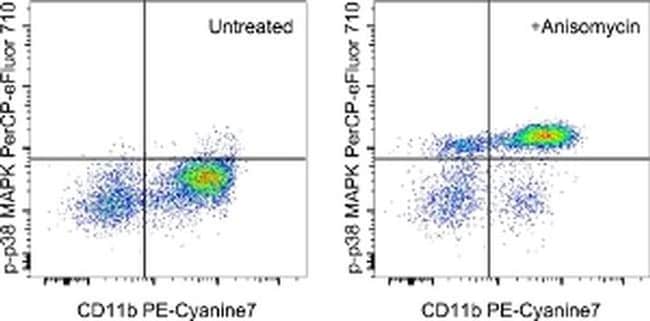
Description: This 4NIT4KK monoclonal antibody recognizes human and mouse p38 mitogen-activated protein kinase (MAPK) when phosphorylated on T180/Y182. p38 MAPK belongs to a family of conserved serine/threonine protein kinases that are phosphorylated and activated in response to numerous stress stimuli including TLR ligands (such as LPS), osmotic shock, heat shock, UV irradiation, and inflammatory cytokines. There are four p38 MAPK members that include p38 alpha, p38 beta, p38 gamma, and p38 delta. The primary activators of p38 MAPK are MKK3/4 and MKK6. Several downstream targets of p38 MAPK have been identified including MK2/3, p53, ATF-2, and ETS1. p38 MAPK can be negatively regulated by the chemical inhibitor SB203580. Specificity of this 4NIT4KK clone was determined by ELISA, flow cytometry, and western blotting. Applications Reported: This 4NIT4KK antibody has been reported for use in intracellular staining followed by flow cytometric analysis. Applications Tested: This 4NIT4KK antibody has been pre-titrated and tested by intracellular staining followed by flow cytometric analysis of stimulated normal human peripheral blood cells. This can be used at 5 μL (0.03 μg) per test. A test is defined as the amount (μg) of antibody that will stain a cell sample in a final volume of 100 μL. Cell number should be determined empirically but can range from 10^5 to 10^8 cells/test.
Specifications
Specifications
| Antigen | Phospho-p38 MAPK (Thr180, Tyr182) |
| Applications | Flow Cytometry |
| Classification | Monoclonal |
| Clone | 4NIT4KK |
| Concentration | 5 μL/Test |
| Conjugate | PerCP-eFluor 710 |
| Formulation | PBS with BSA and 0.09% sodium azide; pH 7.2 |
| Gene | MAPK14 |
| Gene Accession No. | P47811, Q16539 |
| Gene Alias | Crk1; CSAID-binding protein; Csaids binding protein; CSBP; CSBP1; CSBP2; CSPB1; cytokine suppressive anti-inflammatory drug binding protein; cytokine suppressive anti-inflammatory drug binding protein 1; cytokine suppressive anti-inflammatory drug-binding protein; cytokine-supressive anti-inflammatory drug binding protein; Exip; Hog; MAP kinase 11; MAP kinase 13; MAP kinase 14; MAP kinase 2; MAP kinase Mxi2; MAP kinase p38 alpha; MAP kinase p38 beta; MAP kinase p38 delta; MAPK 11; MAPK 13; MAPK 14; Mapk11; Mapk13; MAPK-13; Mapk14; MAX-interacting protein 2; mitogen activated protein kinase 11; mitogen activated protein kinase 13; mitogen activated protein kinase 14; mitogen-activated protein kinase 11; Mitogen-activated protein kinase 13; mitogen-activated protein kinase 14; mitogen-activated protein kinase 14A; mitogen-activated protein kinase p38 alpha; Mitogen-activated protein kinase p38 beta; mitogen-activated protein kinase p38 delta; mitogen-activated protein kinase p38-2; MXI2; p38; p38 alpha; p38 delta MAP kinase; p38 MAP kinase; p38 MAP kinase alpha; p38 MAPK; p38 mitogen activated protein kinase; p38-2; p38a; p38ALPHA; p38-alpha; p38alpha Exip; P38b; p38Beta; p38beta2; p38delta; p38Hog; p38MAPK; Prkm11; PRKM13; PRKM14; Prkm15; protein kinase, mitogen activated kinase, 11, p38beta; reactive kinase; RK; SAPK/Erk/kinase 4; SAPK2; SAPK2A; SAPK2B; SAPK4; Serk4; Stress-activated protein kinase 2a; Stress-activated protein kinase 2b; Stress-activated protein kinase 4; stress-activated protein kinase-2; stress-activated protein kinase-2b; tRNA synthetase cofactor p38 |
| Show More |
Frequently Asked Questions (FAQs)
Our options will depend on the samples you are analyzing.
If cell viability is not critical, you can store your stained samples at 4 degrees C or on ice overnight in the dark and analyze the following day.
For samples stained with eFluor organic fluorochromes, we recommend that cells be suspended in 100 uL of Flow Cytometry Staining Buffer (Cat. No. 00-4222) and 100 uL of eBioscience IC Fixation Buffer (Cat. No. 00-8222); samples can be incubated for up to 3 days at 4 degrees C in the dark. Alternatively, the 1-step Fix/Lyse Solution (Cat. No. 00-5333) can be used. This is a great option when working with whole blood but also works for other cell types.
Yes, the eFluor Organic fluorochromes can be used for intracellular staining. The eFluor organic fluorochromes maintain bright signal and require minimal changes in compensation when fixed with eBioscience IC Fixation Buffer (Cat. No. 00-8222-49) and Permeabilization Buffer (Cat. No. 00-8333-56) or 1-step Fix/Lyse Solution (Cat. No. 00-5333-54, 00-5333-57) (as compared to live cells).
Antibodies are tested and validated in a variety of ways. First, we verify that in a panel of cells in which different pathways have been induced, we see phosphorylation specific staining only in the cells in which the specific pathway of interest has been activated. Second, we verify that phosphorylation specific staining is observed only in cell types in which the protein is expressed and not in cell types in which the protein is not expressed. Third, whenever possible, western immunoblotting is used to confirm the presence of a band(s) of the appropriate size(s) in stimulated/treated cells (and not in unstimulated/untreated cells, as appropriate).
The eFluor Organic fluorochromes and eVolve QDots can be used with flow staining buffers containing PBS and protein.
The eFluor Organic Dyes (eFluor 450, APC-eFluor 780, PerCP-eFluor 710, eFluor 710) are conventional fluorochromes. In contrast, the eVolve line of products are Quantum dots.
For Research Use Only.
By clicking Submit, you acknowledge that you may be contacted by Fisher Scientific in regards to the feedback you have provided in this form. We will not share your information for any other purposes. All contact information provided shall also be maintained in accordance with our Privacy Policy.