Learn More
Invitrogen™ Phospho-SRC (Tyr418) Monoclonal Antibody (SC1T2M3), eBioscience™, Invitrogen™


Description
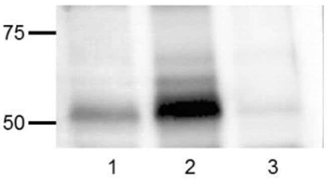
This SC1T2M3 MAb recognizes human and mouse Src tyrosine kinase (also known as ASV, c-src, c-SRC, p60-Src, pp60c-src, Proto-oncogene c-Src, Proto-oncogene tyrosine-protein kinase Src, SRC1) when phosphorylated on tyrosine 418 (Y418). Autophosphorylation of Src at Y418 in the catalytic domain is required for full catalytic activity of this kinase. Src is a non-receptor tyrosine kinase involved in signal transduction in numerous biological systems and is activated following engagement of many different classes of cellular receptors including immune response receptors, integrins and other adhesion receptors, receptor protein tyrosine kinases, G protein-coupled receptors as well as cytokine receptors. Aberrant Src activity has been implicated in the development of numerous types of cancer. Due to the sequence homology surrounding Src Y418, this SC1T2M3 clone is predicted to cross-react with many Src family kinases including Src, Lck, Fyn, and Lyn. Specificity of this SC1T2M3 clone was determined by ELISA, flow cytometry, and western blotting.
Specifications
Specifications
| Antigen | Phospho-SRC (Tyr418) |
| Applications | Western Blot |
| Classification | Monoclonal |
| Clone | SC1T2M3 |
| Concentration | 0.5 mg/mL |
| Conjugate | Unconjugated |
| Formulation | PBS with 0.09% sodium azide; pH 7.2 |
| Gene | SRC |
| Gene Accession No. | P05480, P12931 |
| Gene Alias | ASV; AW259666; csrc; c-SRC; c-src protein, C terminus; EC 2.7.10.2; fc54g04; Neuronal proto-oncogene tyrosine-protein kinase Src; non-tyrosine protein kinase; ORF; OTTHUMP00000174476; OTTHUMP00000174477; OTTHUMP00000174478; p60-Src; p60-Src-1; PP60C-SCR; pp60c-src; pp60src; pp60v-src; protein-tyrosine kinase; proto-oncogene c-Src; protooncogene SRC, Rous sarcoma; Proto-oncogene tyrosine-protein kinase Src; Rous sarcoma oncogene; RP5-823N20.1; sb:cb864; SDR; src; src downstream region; src oncogene; SRC proto-oncogene, non-receptor tyrosine kinase; SRC proto-oncogene, non-receptor tyrosine kinase L homeolog; SRC proto-oncogene, non-receptor tyrosine kinase S homeolog; src.L; src.S; SRC1; src-1; src1-A; src-a; src-b; THC6; transforming protein; tyrosine kinase pp60c-src; tyrosine protein kinase c-src; tyrosine protein kinase pp60-c-src; tyrosine-protein kinase SRC-1; Unknown; v-src avian sarcoma (Schmidt-Ruppin A-2) viral oncogene homolog; v-src sarcoma (Schmidt-Ruppin A-2) viral oncogene homolog; v-src sarcoma viral oncogene; wu:fc54g04; XELAEV_18046582mg; xsrc |
| Show More |
Frequently Asked Questions (FAQs)
Antibodies are tested and validated in a variety of ways. First, we verify that in a panel of cells in which different pathways have been induced, we see phosphorylation specific staining only in the cells in which the specific pathway of interest has been activated. Second, we verify that phosphorylation specific staining is observed only in cell types in which the protein is expressed and not in cell types in which the protein is not expressed. Third, whenever possible, western immunoblotting is used to confirm the presence of a band(s) of the appropriate size(s) in stimulated/treated cells (and not in unstimulated/untreated cells, as appropriate).
We have data demonstrating that fixed cells stored in methanol are stable at -20 degrees C or at -80 degrees C for several weeks. We do not recommend storing fixed cells in eBioscience Foxp3/Transcription Factor Buffer. However, cells fixed with eBioscience Foxp3/Transcription Factor Buffer or eBioscience Intracellular (IC) fixation buffer can be stored in eBioscience IC fixation buffer for up to 3 days at 4 degrees C in the dark.
Note: Please be aware that higher compensation values may be seen with tandem dyes if any other fixative is used apart from eBioscience IC fixation buffer, for 3-day storage.
Each antibody has been tested in three different buffer systems: IC fixation and permeabilization, Foxp3/Transcription Factor Buffer, and IC fixation Buffer/Methanol. The recommended buffer system(s) will be noted on the Technical Data Sheet for the specific antibody.
Because methanol can destroy the epitope recognized by some antibodies, ideally surface staining would be performed at the beginning of the protocol with a methanol-resistant fluorochrome (like eFluor 450, FITC, and eFluor 660).
Please recognize that surface staining before stimulation can have undesired effects due to activation of the cell caused by antibody binding. We recommend staining after stimulation/treatment, fixation, and methanol treatment.
In general, you can use other companies' buffer systems provided that their buffers are of a similar composition as the buffers recommended. Be aware that results will vary depending upon the buffer system used. eBioscience antibodies have been optimized for use in eBioscience buffers and we have not tested all of our antibodies in other buffer systems. Thus, we highly recommend using our optimized protocol and buffer systems.
By clicking Submit, you acknowledge that you may be contacted by Fisher Scientific in regards to the feedback you have provided in this form. We will not share your information for any other purposes. All contact information provided shall also be maintained in accordance with our Privacy Policy.