Learn More
Invitrogen™ Phospho-SRC (Tyr418) Monoclonal Antibody (SC1T2M3), PerCP-eFluor™ 710, eBioscience™


Description
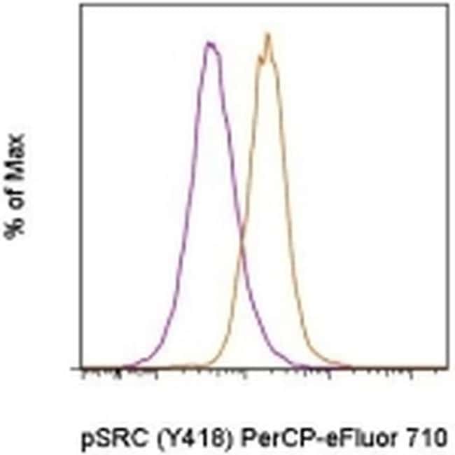
Description: This SC1T2M3 monoclonal antibody recognizes human and mouse Src tyrosine kinase (also known as ASV, c-src, c-SRC, p60-Src, pp60c-src, Proto-oncogene c-Src, Proto-oncogene tyrosine-protein kinase Src, SRC1) when phosphorylated on tyrosine 418 (Y418). Autophosphorylation of Src at Y418 in the catalytic domain is required for full catalytic activity of this kinase. Src is a non-receptor tyrosine kinase involved in signal transduction in numerous biological systems and is activated following engagement of many different classes of cellular receptors including immune response receptors, integrins and other adhesion receptors, receptor protein tyrosine kinases, G protein-coupled receptors as well as cytokine receptors. Aberrant Src activity has been implicated in the development of numerous types of cancer. Due to the sequence homology surrounding Src Y418, this SC1T2M3 clone is predicted to cross-react with many Src family kinases including Src, Lck, Fyn, and Lyn. Specificity of this SC1T2M3 clone was determined by ELISA, flow cytometry, and western blotting. Applications Reported: This SC1T2M3 antibody has been reported for use in intracellular staining followed by flow cytometric analysis. Applications Tested: This SC1T2M3 antibody has been pre-titrated and tested by intracellular staining followed by flow cytometric analysis of normal human peripheral blood cells. This can be used at 5 μL (0.125 μg) per test. A test is defined as the amount (μg) of anti...
Specifications
Specifications
| Antigen | Phospho-SRC (Tyr418) |
| Applications | Flow Cytometry |
| Classification | Monoclonal |
| Clone | SC1T2M3 |
| Concentration | 5 μL/Test |
| Conjugate | PerCP-eFluor 710 |
| Formulation | PBS with BSA and 0.09% sodium azide; pH 7.2 |
| Gene | SRC |
| Gene Accession No. | P05480, P12931 |
| Gene Alias | ASV; AW259666; csrc; c-SRC; c-src protein, C terminus; EC 2.7.10.2; fc54g04; Neuronal proto-oncogene tyrosine-protein kinase Src; non-tyrosine protein kinase; ORF; OTTHUMP00000174476; OTTHUMP00000174477; OTTHUMP00000174478; p60-Src; p60-Src-1; PP60C-SCR; pp60c-src; pp60src; pp60v-src; protein-tyrosine kinase; proto-oncogene c-Src; protooncogene SRC, Rous sarcoma; Proto-oncogene tyrosine-protein kinase Src; Rous sarcoma oncogene; RP5-823N20.1; sb:cb864; SDR; src; src downstream region; src oncogene; SRC proto-oncogene, non-receptor tyrosine kinase; SRC proto-oncogene, non-receptor tyrosine kinase L homeolog; SRC proto-oncogene, non-receptor tyrosine kinase S homeolog; src.L; src.S; SRC1; src-1; src1-A; src-a; src-b; THC6; transforming protein; tyrosine kinase pp60c-src; tyrosine protein kinase c-src; tyrosine protein kinase pp60-c-src; tyrosine-protein kinase SRC-1; Unknown; v-src avian sarcoma (Schmidt-Ruppin A-2) viral oncogene homolog; v-src sarcoma (Schmidt-Ruppin A-2) viral oncogene homolog; v-src sarcoma viral oncogene; wu:fc54g04; XELAEV_18046582mg; xsrc |
| Show More |
Frequently Asked Questions (FAQs)
Our options will depend on the samples you are analyzing.
If cell viability is not critical, you can store your stained samples at 4 degrees C or on ice overnight in the dark and analyze the following day.
For samples stained with eFluor organic fluorochromes, we recommend that cells be suspended in 100 uL of Flow Cytometry Staining Buffer (Cat. No. 00-4222) and 100 uL of eBioscience IC Fixation Buffer (Cat. No. 00-8222); samples can be incubated for up to 3 days at 4 degrees C in the dark. Alternatively, the 1-step Fix/Lyse Solution (Cat. No. 00-5333) can be used. This is a great option when working with whole blood but also works for other cell types.
Yes, the eFluor Organic fluorochromes can be used for intracellular staining. The eFluor organic fluorochromes maintain bright signal and require minimal changes in compensation when fixed with eBioscience IC Fixation Buffer (Cat. No. 00-8222-49) and Permeabilization Buffer (Cat. No. 00-8333-56) or 1-step Fix/Lyse Solution (Cat. No. 00-5333-54, 00-5333-57) (as compared to live cells).
Antibodies are tested and validated in a variety of ways. First, we verify that in a panel of cells in which different pathways have been induced, we see phosphorylation specific staining only in the cells in which the specific pathway of interest has been activated. Second, we verify that phosphorylation specific staining is observed only in cell types in which the protein is expressed and not in cell types in which the protein is not expressed. Third, whenever possible, western immunoblotting is used to confirm the presence of a band(s) of the appropriate size(s) in stimulated/treated cells (and not in unstimulated/untreated cells, as appropriate).
The eFluor Organic fluorochromes and eVolve QDots can be used with flow staining buffers containing PBS and protein.
The eFluor Organic Dyes (eFluor 450, APC-eFluor 780, PerCP-eFluor 710, eFluor 710) are conventional fluorochromes. In contrast, the eVolve line of products are Quantum dots.
By clicking Submit, you acknowledge that you may be contacted by Fisher Scientific in regards to the feedback you have provided in this form. We will not share your information for any other purposes. All contact information provided shall also be maintained in accordance with our Privacy Policy.