Learn More

Invitrogen™ Podoplanin Monoclonal Antibody (eBio8.1.1 (8.1.1)), eFluor™ 660, eBioscience™, Invitrogen™


Description
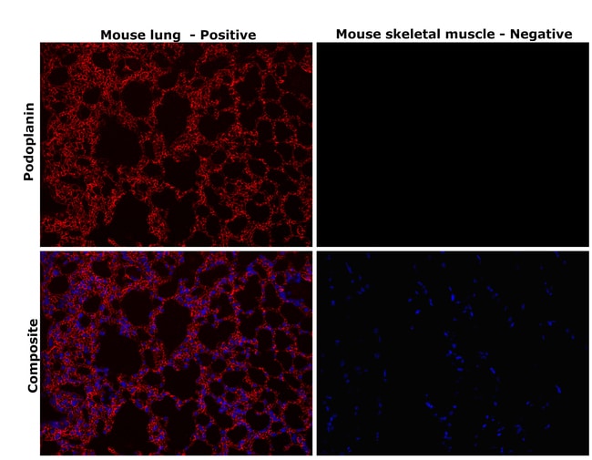
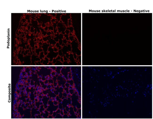
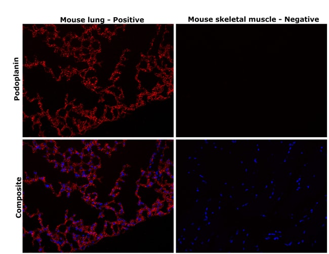
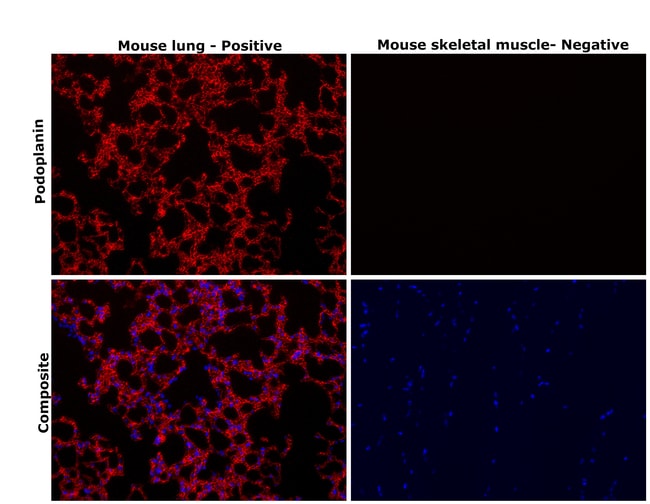
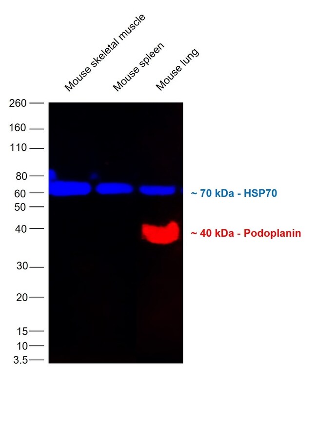
Description: The 8.1.1 monoclonal antibody reacts with mouse podoplanin (T1a, gp38, aggrus), a 43 kDa transmembrane glycoprotein, named for its expression in kidney glomerular epithelial cells (podocytes). In addition, Podoplanin is expressed in epithelial and mesothelial cells such as intestinal epithelium, alveolar type I cells, podocytes, and mesothelium of the visceral peritoneum. It was also shown to be a potent marker for lymphatic endothelium. Podoplanin is also expressed by subcapsular epithelial cells of the murine thymus. Mice deficient in Podoplanin die at birth because of a respiratory defect and congenital lymphedema due to a failure in lymphatic pattern formation. Applications Reported: This eBio8.1.1 (8.1.1) antibody has been reported for use in flow cytometric analysis. Applications Tested: This eBio8.1.1 (8.1.1) antibody has been tested by flow cytometric analysis of the TE-71 cell line. This can be used at less than or equal to 0.125 μg per test. A test is defined as the amount (μg) of antibody that will stain a cell sample in a final volume of 100 μL. Cell number should be determined empirically but can range from 10^5 to 10^8 cells/test. It is recommended that the antibody be carefully titrated for optimal performance in the assay of interest. eFluor™ 660 is a replacement for Alexa Fluor™ 647. eFluor™ 660 emits at 659 nm and is excited with the red laser (633 nm).
Specifications
Specifications
| Antigen | Podoplanin |
| Applications | Flow Cytometry, Immunohistochemistry (Paraffin), Western Blot |
| Classification | Monoclonal |
| Clone | eBio8.1.1 (8.1.1) |
| Concentration | 0.2 mg/mL |
| Conjugate | eFluor 660 |
| Formulation | PBS with 0.09% sodium azide; pH 7.2 |
| Gene | PDPN |
| Gene Accession No. | Q62011 |
| Gene Alias | 29kDa cytosolic podoplanin intracellular domain; Aggrus; CTA-520D8.1; E11; E11 antigen epitope; epithelial cell surface transmembrane protein antigen; glycoprotein 36; Glycoprotein 38; glycoprotein, 36-KD; GP36; Gp38; GP40; HT1A-1; hT1alpha-1; hT1alpha-2; lung type I cell membrane associated glycoprotein; lung type-I cell membrane-associated glycoprotein (T1A-2); Ots8; OTS-8; OTTHUMP00000009640; OTTHUMP00000044504; PA2.26; PA2.26 antigen; PDPN; PICD; Podoplanin; PSEC0003; PSEC0025; pulmonary type I alveolar epithelial cell transmembrane differentiation marker; RANDAM-2; RP23-348F1.2; RTI140; RTI40; T1a; T1A2; T1A-2; T1alpha; T1-alpha; TI1A; Transmembrane glycoprotein E11; type I cell 40 kDa protein |
| Show More |
Frequently Asked Questions (FAQs)
Our options will depend on the samples you are analyzing.
If cell viability is not critical, you can store your stained samples at 4 degrees C or on ice overnight in the dark and analyze the following day.
For samples stained with eFluor organic fluorochromes, we recommend that cells be suspended in 100 uL of Flow Cytometry Staining Buffer (Cat. No. 00-4222) and 100 uL of eBioscience IC Fixation Buffer (Cat. No. 00-8222); samples can be incubated for up to 3 days at 4 degrees C in the dark. Alternatively, the 1-step Fix/Lyse Solution (Cat. No. 00-5333) can be used. This is a great option when working with whole blood but also works for other cell types.
Yes, the eFluor Organic fluorochromes can be used for intracellular staining. The eFluor organic fluorochromes maintain bright signal and require minimal changes in compensation when fixed with eBioscience IC Fixation Buffer (Cat. No. 00-8222-49) and Permeabilization Buffer (Cat. No. 00-8333-56) or 1-step Fix/Lyse Solution (Cat. No. 00-5333-54, 00-5333-57) (as compared to live cells).
Yes, in-house studies have demonstrated that the eFluor 660 fluorochrome is recognized by Anti-Cy5/Alexa Fluor 647 beads. Side by side studies with Alexa Fluor 647 versus eFluor 660 conjugated antibodies have demonstrated comparable results.
The eFluor Organic fluorochromes and eVolve QDots can be used with flow staining buffers containing PBS and protein.
As with other fluorochromes, we recommend minimal exposure to light to maintain optimal signal.
For Research Use Only.
By clicking Submit, you acknowledge that you may be contacted by Fisher Scientific in regards to the feedback you have provided in this form. We will not share your information for any other purposes. All contact information provided shall also be maintained in accordance with our Privacy Policy.