
Microscope Accessories and Replacement Parts
General accessories and parts for use with microscopes. Products include lens oil, lens cleaner, slide dispensers, microscope covers, vessel holders, light cubes, and flow cells.
Many parts of your microscope can be updated or replaced including microscope lamps, lenses, and eyepieces. Replacement lamps, light sources, and power cords can extend the working lives of your microscopy equipment. New or replacement objectives and lenses can help increase the total magnification available with your microscope.
Add greater magnification or exchange your eyepiece for one with a reticule to more easily estimate the sizes of the items being viewed.
Replace or update your mechanical stage or clamps or add cell culture vessel adapters to facilitate specimen positioning.
Condensers can also be replaced, and conversion kits are available to add phase contrast, polarization, fluorescence, or other capabilities to your current system.
Some microscopes can be adapted for use with still or video cameras. Look for cameras and adapters designed for your specific microscope brand. Film and documentation-related products may also be available to support your record-keeping.
Temporary, semi-permanent, and more long-term slide storage options include:
- Slide racks that securely hold slides during staining procedures
- Folding fiberboard or plastic slide holders with horizontal indentations that keep microscope slides in place
- Slide storage boxes with vertical dividers; made from fiberboard, plastic, and wood and available in multiple sizes
- Slide holders and mailers for transporting slides safely
- Slide storage systems and cabinets for large quantities of slides or archival storage

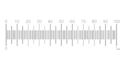
Electron Microscopy Sciences PS8 Micrometer Standard with NPL Cert
These stage micrometers are available with a calibration certificate that provides traceability for the precise calibration and confirmation of accuracy of optical measuring instruments, necessary under ISO provisions. The glass discs are mounted in stainless steel slides with engraved serial numbers. Each slide is supplied in a polished wooden storage case. Calibrated by the National Physical Laboratory by taking measurements at selected points on the scale.


Electron Microscopy Sciences Gilder Standard Square 300 mesh Cu/Pd Grid
A reliable support specimen grid source. Features well-defined grid bars, maximum open area, and a matt/shiny side. Each grid is individually inspected. 3.05 mm diameter, 0.7 mil (18 um) thickness.
Grid: Copper/Palladium
Pitch: 83 µm
Hole: 58 µm
Bar: 25 µm
:
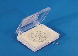
Electron Microscopy Sciences BEEM Dial-A-Grid Storage Box
A two tone color coded plastic box with insert which has 24 letter-labeled crossed slots, where the grids can be stored. A rotating protection plate covers the slots and allows for exposure of one grid at a time. BEEM Is A Registered Trademark of Better Equipment For Electron Microscopy, Inc.

Leica Biosystems HC PL FLUOTAR 20x/0.55 Microscope Objective (20x, NA 0.55)
HC PL FLUOTAR 20x/0.55 is a fluorescence-optimized microscope objective that provides 20x magnification with a 0.55 numerical aperture. It delivers flat-field correction and chromatic control suitable for fluorescence and transmitted-light imaging in routine and research microscopy workflows.
- 20x magnification with 0.55 numerical aperture.
- Fluorescence-optimized optical design for high-contrast imaging.
- Plan correction for a flat, well-corrected field of view.
- Appropriate for routine and advanced light-microscopy applications.
- Compatible with standard microscope objective turrets and parfocal systems.

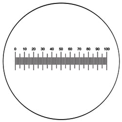
Electron Microscopy Sciences Microscope Graticule NE1 Horizontal, 19 mm, 10 mm with 0.1 mm subdivisions
These scales are used for the measuring of lengths of specimen or distances between points on a variety of different shaped objects. By dividing the division of the chosen graticule by the magnification of the objective one obtains an approximate value that each division will represent on the stage. Horizontal micrometer scale.
Size: 19 mm
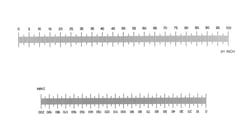
Electron Microscopy Sciences Stage Micrometer S20, Double micrometer scale Scale, Length
Stage graticules are used for the routine calibration of eyepiece patterns, particularly when alternating between objectives on one microscope or when using the same graticule in different microscopes. Their robust construction makes them ideal for student use and for instructional purposes. The scale or grid is centered on a glass disc mounted in a black anodized aluminum slide 75 mm x 24 mm x 2 mm thick.
Scale: Double micrometer scale
Divisions: 2 mm in 0.01 mm and 0.1"in 0.005"

Electron Microscopy Sciences Gilder Thin Bar Square 2000 mesh Nickel Grid
Very fine mesh grids have up to 2,000 lines/inch. There is an increasing need in TEM for support films, routinely carbon, as thin as 1.5 - 2.0 nm. The pitch (the distance from the center of one bar to the center of the next bar) dimension in all grids remains constant, allowing use as a lower magnification calibration aid.
Apart from one side being shiny, the other matt, there is a large asymmetrical mark in the rim which gives the identification of which side the specimen is on. Grids are 3.05 mm overall diameter; 2.05 mm mesh area diameter.
Grid: Nickel
Pitch: 12.5 µm
Hole: 7.5 µm
Bar: 5 µm
:

Electron Microscopy Sciences TEM Specimen Supports Molybdenum GS2 x 1
We have extended our range of TEM grid materials to include four types, which are now available in Molybdenum. The new products are manufactured using a process known as chemical ʽmilling' (etching) instead of the more familiar technique of ʽelectroforming' (deposition) that is used in the manufacture of copper, nickel and gold products. Molybdenum is used principally in applications where it's high temperature, hardness, expansion of coefficient and corrosion resistance characteristics are considered important. The material which is used has a purity of 99.9%.
Hole: 2 x 1 mm
Thickness: 50 µm

Electron Microscopy Sciences V-2 Quality, 25 x 75 mm, 0.26- 0.31 mm thick
EMS Mica Sheets offer a clean surface for EM applications, carbon filming and particle spraying, as well as for AFM applications. There are two types of mica: muscovite and phlogopite. Generally, one differs from the other by color (Muscovite is Ruby, Green or White; Phlogopite is Amber, Yellow, or Silver). The maximum operating temperature for Muscovite is about 500-600 degree C and for Phlogopite is about 800-900 degree C. Our line consists of High Quality Muscovite Mica. This mica peels off very thin up to 0.0001" uniform layers, exposing "virgin" mica upon splitting.Muscovite, potash type mica, sometimes known as granitic mica, is the best of all micas in dielectric strength, perfection of cleavage, and transparency. It has the chemical formula: H2KAI3(Si04)3. Clear and Slightly Stained. Hard, uniform color, nearly flat and slight crystallographic discoloration. Quality: V-2Size: 25 x 75 mmThickness: 0.26- 0.31 mm

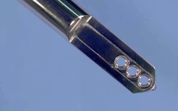
Electron Microscopy Sciences Hitachi 3-Position Multi-Sample Holder
A single tilt multi-grid holder. Will accommodate 3 grids with easy reproducible movement between each specimen. It is ideal for scanning through serial samples or for comparing against standards. Grids are held in place by easy to remove circlips. Comes complete with loading stand, circlip extraction tool, and is supplied in a mahogany box.
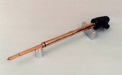
Electron Microscopy Sciences JEOL 2-Position Single Tilt Holder
A single tilt multi-grid holder. Will accommodate 6 grids with an easy click stop reproducible movement between each specimen. It is ideal for scanning through serial sections or for comparing against a standard. Grids are held in place by easy to remove circlips. Comes complete with loading stand, circlip extraction tool, and is supplied in its own mahogany box.
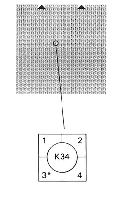
Electron Microscopy Sciences S7 England Finder Slide
S7 is a glass slide 75 mm x 26 mm marked over the top surface in a way that a referenced position can be directly read relative to the locating edges. It is marked with a square grid at 1 mm intervals. Each square contains a center ring bearing reference letters and numbers, the remainder of the square is subdivided into four segments numbered 1 to 4.
Reference numbers run horizontally 1 to 75 and letters vertically A to Z (omitting I). The main locating edge is the bottom of the slide, which is used in conjunction with either the left or right vertical edge of the slide.
Using the finder the position of a field of interest is easily recorded, ite can be relocated by another person when using a England Finder on another microscope. The location of the arrows is identical for all England Finder slides. Mark a label on the left hand side of the specimen slide, indicating the orientation. A total of 100 positions can be accurately located.
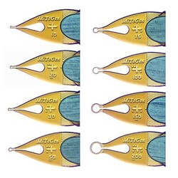
Electron Microscopy Sciences MicroMounts™ Crystal Tool with 10 µm Aperture and SPINE pin
MicroMounts are the high performance tools for manipulating and mounting protein crystals, virus crystals, and small molecule/inorganic crystals, and other small, fragile samples. They are ideal for both conventional and high-throughput X-ray crystallography applications.
Each consist of a thin (7.5um thick) micro-fabricated polyimide film, attached to a solid non-magnetic stainless steel pin. Film is wrapped around the steel pin and it provides excellent stiffness and convenient scoop-like action in retrieving and handling crystals.
Aperture: SPINE pin