
Protein Labeling Reagents


Medchemexpress LLC Fosbretabulin disodium | 168555-66-6 | 99.9% | 440.29 | 5 MG

Fosbretabulin disodium (CA 4DP) is a tubulin destabilizing agent. It functions as a Combretastatin A4 proagent, selectively targeting endothelial cells. This action induces regression of nascent tumor neovessels, reduces tumor blood flow, and causes central tumor necrosis. It has demonstrated efficacy in inhibiting the growth of various cancer cell lines in vitro and reducing tumor blood flow in animal models.
- Acts as a tubulin destabilizing agent.
- Selectively targets endothelial cells.
- Induces regression of nascent tumor neovessels.
- Reduces tumor blood flow.
- Causes central tumor necrosis.
- Inhibits growth of multiple cancer cell lines.
- Decreases tumor blood flow in animal models.


Biotium p53(PAb122), CF568 conjugate, 0.1mg/mL
Mutation and/or allelic loss of p53 is one of the causes of a variety of mesenchymal and epithelial tumors. If it occurs in the germ line, such tumors run in families. p53 Binds to a DNA consensus sequence, the p53 response element, and it regulates normal cell growth cycle events by activating transcription of genes, involved either in progression through the cycle, or causing arrest in G1 when the genome is damaged. In most transformed and tumor cells the concentration of p53 is increased 51000 fold over the minute concentrations (1000 molecules cell) in normal cells, principally due to the increased half-life (4 h) compared to that of the wild-type (20 min). p53 Localizes in the nucleus, but is detectable at the plasma membrane during mitosis and when certain mutations modulate cytoplasmic/nuclear distribution. p53 Is the most commonly mutated gene in spontaneously occurring human cancers. Mutations arise with an average frequency of 70% but incidence varies from zero in c
Biotium CD45RO(UCHL-1), CF568 conjugate, 0.1mg/mL
Recognizes a 180-185 kDa protein, identified as isoform of leukocyte common antigen (CD45RO) (4th Leucocyte Typing Workshop: Code No. N31). The epitope recognized by this antibody is sensitive to neuraminidase digestion. This antibody reacts with mature activated T-cells, most thymocytes, and a sub-population of resting T-cells within both CD4 and CD8 subsets. It shows no reactivity with normal B or natural killer cells, but reacts with granulocytes and monocytes. Reportedly, it is useful to identify T-cell lymphomas and leukemias. It rarely stains NK cells or B-cell lymphomas.Primary antibodies are available purified, or with a selection of fluorescent CF Dyes and other labels. CF Dyes offer exceptional brightness and photostability. Note: Conjugates of blue fluorescent dyes like CF405S and CF405M are not recommended for detecting low abundance targets, because blue dyes have lower fluorescence and can give higher non-specific background than other dye colors.
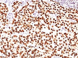
Biotium PLGF(PLGF93), CF568 conjugate, 0.1mg/mL
The onset of angiogenesis is believed to be an early event in tumorigenesis and may facilitate tumor progression and metastasis. Several growth factors with angiogenic activity have been described. These include Fibroblast Growth Factor (FGF), Platelet Derived Growth Factor (PDGF), Vascular Endothelial Growth Factor (VEGF) and Placenta Growth Factor (PLGF). Placenta growth factor (PLGF) is a secreted protein primarily produced by placental trophoblasts but also expressed in other endothelial cells and tumors. There are three isoforms, PLGF-1, PLGF-2, and PLGF-3. PLGF-2 is expressed up until week 8 in the placenta; the placental tissues continuously express PLGF-1 and PLGF-3 but only PLGF-1 is found in colon and mammary carcinomas. PLGF acts to stimulate angiogenesis, endothelial growth and migration. PLGF is a powerful promoter of tumor growth and is upregulated in some cancers, and PLGF is thought to aid in atherosclerotic lesions and neovascular growth surrounding the lesio
Biotium CD36(185-1G2), CF568 conjugate, 0.1mg/mL
Recognizes a protein of 80 kDa-90 kDa, identified as CD36 (Workshop IV; Code P-26). Its epitope maps between aa155-183. It is expressed on platelets, monocytes and macrophages, microvascular endothelial cells, erythrocyte precursors, mammary epithelial cells, and some macrophage derived dendritic cells. CD36 acts as a receptor for thrombospondin (TSP), collagen types I, IV and V, P. falciparum malaria-infected erythrocytes, and sickle erythrocytes. It also functions as a scavenger receptor, mediating macrophage uptake of oxidized low-density lipoprotein (LDL) and recognition of apoptotic polymorphonuclear leukocytes (PMN). CD36 plays a role in platelet aggregation, macrophage foam cell development, inflammation, and the tissue ischemia observed in sickle cell disease and cerebral malaria. Note that 1-4% of Japanese and East Asia population lack CD36. This MAb blocks adhesion of P. falciparum parasitized red blood cells to CD36 and strongly inhibits collagen-induced platelet a
NCCCN=[N+]=[N-])c3ccc(cc3C2(C)C)S([O-])(=O)=O)N(CCCS(O)(=O)=O)c2ccc(cc12)S(O)(=O)=O | 1mg | 268505019](https://assets.fishersci.com/TFS-Assets/CCG/product-images/502465212.JPG-250.jpg)
eMolecules 1801695-56-6 | Sulfo-Cy3 Azide | Broadpharm | MFCD30527437 | 806.970 | C35H46N6O10S3 | 90.000 | CC1(C)\C(=C/C=C/C2=[N+](CCCCCC(=O)NCCCN=[N+]=[N-])c3ccc(cc3C2(C)C)S([O-])(=O)=O)N(CCCS(O)(=O)=O)c2ccc(cc12)S(O)(=O)=O | 1mg | 268505019
Sulfo-Cy3 Azide | Broadpharm | 1801695-56-6 | MFCD30527437 | 806.970 | C35H46N6O10S3 | 90.000 | CC1(C)\C(=C/C=C/C2=[N+](CCCCCC(=O)NCCCN=[N+]=[N-])c3ccc(cc3C2(C)C)S([O-])(=O)=O)N(CCCS(O)(=O)=O)c2ccc(cc12)S(O)(=O)=O | 1mg | 268505019
Biotium Fascin-1(FSCN1/418), CF488A conjugate, 0.1mg/mL
Recognizes a protein of 55 kDa, which is identified as fascin-1. Its actin binding ability is regulated by phosphorylation. Antibody to fascin-1 is a very sensitive marker for Reed-Sternberg cells and variants in nodular sclerosis, mixed cellularity, and lymphocyte depletion Hodgkin's disease. It is uniformly negative in lymphoid cells, plasma cells, and myeloid cells. Fascin-1 is also expressed in dendritic cells. This marker may be helpful to distinguish between Hodgkin lymphoma and non-Hodgkin lymphoma in difficult cases. Also, the lack of expression of fascin-1 in the neoplastic follicles in follicular lymphoma may be helpful in distinguishing these lymphomas from reactive follicular hyperplasia in which the number of follicular dendritic cells is normal or increased. Antibody to fascin-1 has been suggested as a prognostic marker in neuroendocrine neoplasms of the lung as well as in ovarian cancer. Fascin-1 expression may be induced by Epstein-Barr virus (EBV) infection o
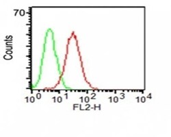
Biotium CD34(ICO-115), CF568 conjugate, 0.1mg/mL
This antibody recognizes a carbohydrate epitope on a single chain, transmembrane, heavily glycosylated protein of 90-120 kDa, which is identified as CD34 (VI international workshop on human differentiation antigens). Its expression is a hallmark for identifying pluripotent hematopoietic stem or progenitor cells. Its expression is gradually lost as lineage committed progenitors differentiate. CD34 is a marker of choice for staining blasts in acute myeloid leukemia. In addition, it is expressed by soft tissue tumors, such as solitary fibrous tumor and gastrointestinal stromal tumor. CD34 expression is also found in vascular endothelium. Additionally, proliferating endothelial cells overexpress this molecule than the non-proliferating endothelial cells. Anti-CD34 labels > 85% of angiosarcoma and Kaposi's sarcoma, but shows low specificity.Primary antibodies are available purified, or with a selection of fluorescent CF Dyes and other labels. CF Dyes offer exceptional brightness
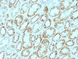
Biotium Laminin(A5), CF488A conjugate, 0.1mg/mL
Laminins are large hetero-trimeric, non-collagenous glycoproteins composed of α, β, and γ chains. This MAb reacts with laminin B2/1 chain of ~210 kDa and does not cross-react with other basement membrane components or fibronectin. Its specificity was established by immunoprecipitation and immunofluorescence of human skeletal muscle and kidney with laminin chain-specific MAbs. Epithelial sheets in vivo are separated from the mesenchymal elements of the stroma by a thin layer of a specialized type of extracellular matrix termed the basement membrane (BM). This structure consists of individual components, some of which are ubiquitous in BMs and some are not. The ubiquitous ones comprise laminin (LN), entactin/nidogen (EN), collagen type IV (CIV), and large heparan sulfate proteoglycan (HSPG), which interact specifically with each other to form a continuous and regular BM. Alterations of BM integrity, from local discontinuities up to complete loss, are describe
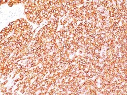
Biotium CD45RB(BRA-11G), CF488A conjugate, 0.1mg/mL
CD45R, also designated CD45 and PTPRC, has been identified as a transmembrane glycoprotein, broadly expressed among hematopoietic cells. Multiple isoforms of CD45R are distributed throughout the immune system according to cell type. These isoforms arise because of alternative splicing of exons 4, 5, and 6. The corresponding protein domains are characterized by the binding of monoclonal antibodies specific for CD45RA (exon 4), CD45RB (exon 5), CD45RC (exon 6) and CD45RO (exons 4 to 6 spliced out). The variation in these isoforms is localized to the extracellular domain of CD45R, while the intracellular domain is conserved. CD45RB is expressed on mature B-lymphocytes and the majority of lymphomas and leukemias of B-cell origin.Primary antibodies are available purified, or with a selection of fluorescent CF Dyes and other labels. CF Dyes offer exceptional brightness and photostability. Note: Conjugates of blue fluorescent dyes like CF405S and CF405M are not recommended for

Lumiprobe Sulfo-Cyanine7.5 NHS ester, 1 mg
Sulfo-Cyanine7.5 is a near infrared water soluble and hydrophilic dye for the NIR imaging applications.
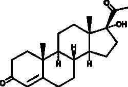
Cayman Chemical 17a-Hydroxyprogesteron 10g
A steroid and precursor in the synthesis of adrenal androgens; binds to the glucocorticoid receptor in a cell-free assay (IC50 = 82 nM for the human receptor) and induces glucocorticoid receptor transactivation in HEK293 cells (EC50 = 1.2 µM); inhibits aldosterone-induced transactivation of the mineralocorticoid receptor in cell-based assays from 0.005-1 µM; serum levels are increased in patients with congenital adrenal hyperplasia
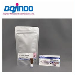
Dojindo Molecular Technologies Inc PlasMem Bright Red dye (100 ul)
The plasma membrane consists of a lipid bilayer separating the intracellular environment from the extracellular space. Thus, the PM plays a central role in many cell behaviors such as cell migration, cell stretching, and signaling cascades. Additionally, PM dysfunction is an important biomarker because it is related to cell status and is linked to many diseases. PlasMem Bright dyes have higher retentivity on the plasma membrane. Furthermore, PlasMem Bright dyes are more water-soluble compared with other commercially available dyes and can be diluted with the culture medium. The PlasMem Bright dyes offer two different color options (green and red) and provide ready-to-use DMSO solutions. A working solution can be prepared easily via a single dilution step using growth medium or HBSS. Dojindo offers various reagents for cell membrane research. (https://www.dojindo.com/track-the-dynamics-of-plasma-membrane/)

Biotium CF568 Hydrazide
Our CF dye hydrazides are bright, extremely water-soluble and nontoxic. These properties make the dyes excellent fixable polar tracers for microinjection into cells. Hydrazides also can be used for labeling aldehyde or ketone groups, however, for this application we recommend using the more reactive CF dye aminooxy forms. CF dyes are Biotium's line of next-generation fluorescent dyes with advantages in brightness, photostability, and conjugate specificity compared to other fluorescent dyes. Red fluorescent CF568 dye has excitation/emission at 562/583 nm.